INTRODUCTION
Transforaminal percutaneous endoscopic lumbar discectomy (PELD) is one of the established surgical modalities for treating symptomatic disc herniations [4,5,19,20]. Postoperative dysthesia or motor weakness from exiting root injury is a unique complication of PELD which usually is caused by the cannula placed in the Kambin’s zone near the exiting nerve root [1,2,4-6,13,19,20]. Transforaminal access involves passing endoscopes and instruments through a cannula placed safely in the Kambin’s safe zone11) (an important landmark) to avoid exiting root injury. The limited dimensions of Kambin’s triangle limit the size of cannula and instruments that can be passed through it.
The study by Mirkovic et al. [17] is the only cadaveric study which calculated that 7.5 mm or 6.3 mm cannula can be used safely depending on the placement of the cannula with respect to the pedicle, while Lertudomphonwanit et al. [14] calculated the trapezoidal intervertebral disc space area which was safe to introduce a cannula and Min et al. [16] calculated the linear measurements of root to facet distance. However, while performing PELD, facet joint plays an important role in limiting the size of the cannula placed between it and the exiting nerve root. Additionally, in cadaver’s formalin induced tissue changes alter the Kambin’s zone dimensions [3,12,15,18]. Magnetic resonance imaging (MRI) studies have an advantage over cadaveric studies in giving more clinically accurate measurements. Choi et al. [6] and Hurday et al. [10] calculated root to facet joint distances in their MRI based studies. Coronal MRI view is routinely used for pre-operative evaluation of exiting roots for PELD. However, since the exiting nerve root travels obliquely in an infero-ventral direction [7], we feel that for an accurate anatomical analysis of the Kambin’s triangle whose anatomical plane is decided by the direction of the exiting root, a 3D MRI would provide us with the adjustments necessary to take oblique sections in the plane of the exiting root. Our 3D MRI based study is an anatomical study, which treated the original Kambin’s triangle as described by Mirkovic et al. [17] and Kambin as the “neural” Kambin's triangle because of its two neural boundaries (exiting nerve root and dural sheath). However, before reaching the neural Kambin’s triangle, the cannula needs to negotiate the region between the facet and exiting nerve root. We described the working triangle in this region as “bony” Kambin’s triangle because of its two bony boundaries (facet joint and superior end plate of inferior vertebra).
Rendering images from 3D MRIs of lumbar spine, we calculated the dimensions of the neural and bony Kambin's triangles. We then calculated the inscribed circle within these triangles which represents the maximum diameter of cannula permissible through them. Our study is the first to analyse the dimensions of the Kambin's zone on 3D MRI of the lumbar spine.
MATERIALS AND METHODS
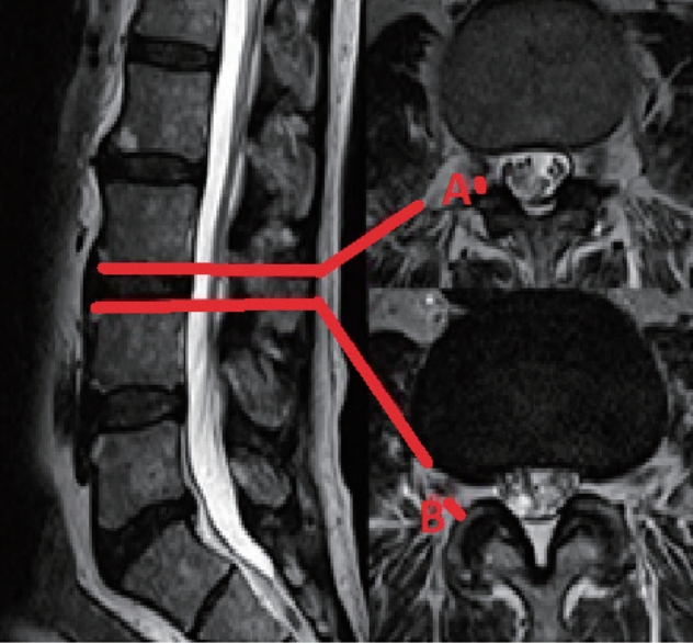
This is an observational prospective 3D MRI based anatomical study. This study protocol was reviewed and approved by Ethics Committee of Deenanath Mangeshkar Hospital where this study was conducted (IRB No. 2015_APR_PP_169). This study being an observational imaging-based study with none of the patients’ identity being disclosed, informed consent was necessary. We studied 3D MRI’s of 50 patients in our study. Patients included were those who presented in Deenanath Mangeshkar Hospital with back pain and/or leg pain between March 2016 and February 2017 and had underwent a 3D MRI of the lumbar in a 3Tesla machine in Deenanath Mangeshkar Hospital. 3D MRI acquisition with reformatting of the images was done of all these patients. Exclusion criteria were fractures, infection, tumour, instability, severe stenosis, previous lumbar surgery, congenital defects and large disc herniation. Disc levels from L12 to L5S1 were evaluated bilaterally. Distances from the exiting nerve root to the closest point on the facet joint at the level of upper and lower end plate of the disc were measured bilaterally taken parallel to the respective end plate on axial sections (Fig. 1).
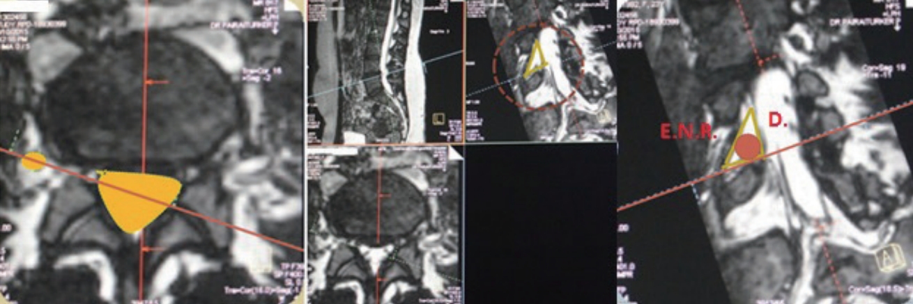
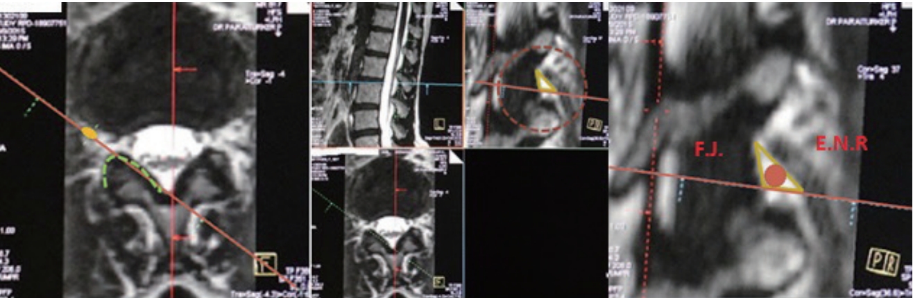
Two sets of anatomical triangles were evaluated on both sides. The neural (original) Kambin’s triangles bound by the exiting root (the hypotenuse), the dura (vertical limb) and the superior end plate of the inferior vertebrae (the base) were evaluated with the axial axis fixed parallel to the lower end plate of the respective disc and the oblique axis adjusted to bisect the exiting nerve root and the dural sheath was taken (Fig. 2). The bony Kambin’s triangles bound by the exiting root (the hypotenuse), the facet i.e., superior articular process of inferior vertebra (the vertical limb) and the superior end plate of the inferior vertebrae (the base) were evaluated with the axial axis fixed parallel to the lower end plate of the respective disc and the oblique axis adjusted to bisect the exiting nerve root and the facet joint was taken (Fig. 3). The diameters of the inscribed circles in both these triangles, which represented the outer diameter of cannula permissible through the neural and bony Kambin’s triangle, were mathematically calculated. First three authors of this study evaluated all readings individually and an average value was taken to reduce errors.
Statistical analysis
All the statistical analyses of the data were processed on a computer by using commercially available SPSS Windows version 19 (SPSS, Chicago, IL, USA). Descriptive statis tics (mean, standard deviation, maximum, and minimum) were calculated. The two-tailed paired t test was used for comparison between the groups. Probability values of p<0.05 considered statistically significant depending on the variable.
RESULTS
There were total 50 patients (19 males and 31 females) with age 18 to 78 years (mean, 47±14). Root to facet distance at upper and lower end plate levels of the disc on both sides from L12 through L5S1 levels were also calculated on axial sections (Fig. 1). The mean root to facet distances at upper end plate level increased from 3.42±3.01 mm at L12 level to 5.13±2.93 mm at L5S1 level (Table 1). The mean root to facet distances at lower end plate level also increased from 6.07±1.13 mm at L12 level to 12.9±2.83 mm at L5S1 level (Table 1). There was no statistically significant difference between right and left sides at each level. Mean maximum permissible endoscopic cannula diameters through the neural Kambin’s triangle gradually increased from L12 to L5S1 level which were 5.67±1.38 mm at L12 level to 9.7±3.82 mm at L5S1 level (Table 2). There was no statistical difference between the right and the left sides. The mean maximum permissible endoscopic cannula diameter for bony Kambin’s triangle also increased from L12 to L5S1 levels which were 4.03±1.08 mm at L12 level to 6.11±1 mm at L5S1 level (Table 2). There was no statistical difference between the right and the left sides.
Out of the total 427 bony Kambin’s triangle evaluated only 2% could accommodate a cannula diameter of 8 mm, 62% could accommodate cannula diameter between 4 to 8 mm and 36% accommodated less than 4 mm cannula diameter (Table 3).
The exiting root to facet distance measured at the lower end plate level measured in axial sections was compared to the base of bony Kambin’s triangle in oblique sections of their respective levels bilaterally. The combined values of both sides were taken and the comparison was done (Table 4). The base of bony Kambin’s triangle was significantly higher at L12, L34, and L45 levels as compared to their respective root to facet distance at lower end plate level, whereas it was not significant at L23 and L5S1 levels (Table 4). Whereas, if the combined measurements of all levels were compared, then the base of bony Kambin’s triangle was significantly higher than the root to facet distance at lower end plate level (p value=0.023) (Table 5).
DISCUSSION
PELD is a well-established procedure [4-6,19,20] with Kambin and Gellman [11] first elaborating the safe anatomical zone, the Kambin’s triangle, for accessing the transforaminal region; which is bound by the exiting root (the hypotenuse), the dura (vertical limb) and the superior end plate of the inferior vertebrae. The boundaries described above limit the size of cannula and the working channel instruments. Until now, most literature evaluating dimensions of Kambin’s triangle have done so by performing cadaveric studies. Mirkovic et al. [17] in their cadaver study, after facetectomies he calculated cannula diameters to be 7.5 mm or 6.3 mm which could be used safely depending on the placement of the cannula with respect to the pedicle. However, we think as the exiting root is in close approximation with the facet joint, it is an important landmark in PELD which needs to be negotiated first before reaching Kambin’s triangle. So, like the original Kambin’s triangle, this triangular region bound by superior articular process (facet joint), the exiting nerve root and lower end plate also limits the size of the cannula placed in it. We referred to this region as the “bony” Kambin's triangle. Among other cadaveric studies, Lertudomphonwanit et al. [14] evaluated the trapezoidal intervertebral disc space area, calculating the largest ellipsoidal cannula that could be placed in IVD area was 5.83×11.02 mm at L1-2, 6.97×10.78 mm at L2-3, 9.30×10.67 mm at L3-4, 8.84 ×13.15 mm at L4-5, and 6.61×14.07 mm at L5-S1. Similarly, Hoshide et al. [9] evaluated surface area of the Kambin’s triangle in his two cadavers. But an endoscopy cannula is not ellipsoid and a triangle's surface area is not representative of the cannula diameter. Min et al. [16] evaluated the root to superior articular process distance, which were from 8.1±2.6 mm at L12 to 15.5±4.2 mm at L5S1 which were higher than those found in our study (6.07±1.31 mm to 12.99±2.83 mm). Güvençer et al. [8] calculated the lateral edge of the root to lateral edge of the superior articular process. But the findings were not comparable to our study, with few measurements being higher and a few being lower. Both these studies [8,16] have measured linear dimensions to evaluate a region which is a 3D structure placed in an oblique plane in relation to the exiting root [7]. We think that although the region between the exiting root and the facet joint has been taken into consideration for pre-operative evaluation before PELD, no cadaveric study has been done to find the actual dimensions of this triangular space (which we have described as bony Kambin’s triangle) or the diameter of largest cannula safely negotiating the region between exiting nerve root and facet joint. Additionally, cadaveric studies have their own limitations e.g., formalin in cadaver preservations causes alterations in tissue tension leading to altered foramina dimensions [3,12,15,18].
MRI as an imaging modality excels in showing the neural structures in great detail and trumps cadaveric studies in giving the best possible clinically usable anatomic evaluation. In his MRI study, Choi et al. [6] calculated the root to facet distance at lower end plate levels which was 6.4±1.5 mm in those without root injury to 4.1±0.8 mm in those with root injury. Hurday et al. [10] evaluated the same distances to be 6.41±1.97 mm at L23 level to -8.53±11.11 mm at L5S1 level. Our measurements were higher than those found in both these studies, with 6.1±1.72 mm at the level of L12 which gradually increased to 12.99±2.83 mm at the level of L5S1 (Table 1). In our study, the root to facet distance at the level of upper end plate of the respective disc was 3.42±3.01 mm at the level of L12 gradually increasing to 5.13±2.93 mm at the level of L5S1. However, the studies of Choi et al. [6] and Hurday et al. [10] provide us with data of linear measurements. As the exiting nerve root travels obliquely in an infero-ventral direction coronal, sagittal or axial images will never give accurate anatomical details to evaluate the size of the circular cannula permissible through the obliquely placed safe zone, which is a 3D structure [7]. Oblique sections, which can be obtained with 3D MRI, provide data which accurately assesses the Kambin’s region. Ours is the first study which did volumetric acquisition of 3D data from MRI’s of the lumbar spine and then by adjusting the obliquity in reformatted images, calculated the dimensions of the bony and neural Kambin’s triangles. On getting the triangle measurements, we mathematically calculated the inscribed circle which represents the largest cannula diameter permissible in these triangular zones.
In our study we found that the mean maximum diameter of endoscopic cannula permissible through the neural Kambin’s could accommodate a 7 mm endoscopic cannula at L34 level with the maximum diameter of 7.52±1.82 mm and an 8 mm endoscopic cannula at L45 and L5S1 levels with the maximum diameters being 8.31±1.98 mm and 9.7±3.82 mm respectively (Table 2). L45 and L5S1 are the most common levels of disc prolapsed requiring PELD. Our MRI based study shows that an 8mm cannula can be easily accommodated at these levels within the original (neural) Kambin’s triangle. However, the maximum diameter of cannula permissible through the bony Kambin’s triangle was highest at L5S1 which was 6.11±1 mm with least being 4.03±1.08 mm at L12 level (Table 3). So, a 7 mm or 8 mm cannula might have difficulty passing between the facet joint and exiting nerve root. Additionally, out of the total 427 bony Kambin’s triangles evaluated, only 2% could accommodate a cannula diameter of 8 mm with 62% accommodating a 4-8 mm cannula and 36% accommodating a 4 mm cannula (Table 2). So there exists a contradiction between the safe sizes cannulas advised for PELD in literature [17] and the observations of our study that a cannula of a maximum 6.11±1 mm diameter can be safely placed within the facet and exiting root. The only reason which can explain placement of an 8 mm cannula between the facet and exiting root is by gradual root retraction.
Choi et al. [6] stated that after PELD the complication rate of exiting nerve injury increased by 23% per each 1-mm decrease in distance from root to facet at lower disc level. However, the reported incidence of exiting nerve root injury during transforaminal discectomy is not very high (1% to 6.7%) [1,2,4-6,19,20]. This means that the established PELD procedures, which include the rail-road technique of introducing a tapered dilator followed by a snugly fitting cannula, don’t cause exiting root injury as frequently [4,19,20] as statistically suggested by Choi et al. [6]. Interestingly, he further reported that those 20 patients with exiting root injury had significantly high operative time as opposed to those who did not, with each minute increase in operative time increases chances of exiting root injury by 1.027-fold. In light of these findings, we need to really evaluate whether the time duration is the major deciding factor for exiting root injury rather than root retraction.
We considered the fact that it may not be possible in all centres to get a pre-operative 3D MRI. The root to facet distance at the lower end plate level measured on axial sections corresponds to the base of the bony Kambin’s triangle as measured on oblique sections in 3D MRI. So, we assumed that if both these would have been same in our study we might do away with the necessity of using a 3D MRI pre-operatively. However, the base of the bony Kambin’s triangle as measured by oblique sections was significantly higher than root to facet distance at the lower end plate level measured on axial sections (Table 5). This discrepancy may be explained by the different points of references used in both different views for measuring distances because of the oblique course of the exiting nerve root. So, it is necessary to use a 3D MRI for pre-operative evaluation of the Kambin’s zone.
There are limitations in our study. Ours is an MRI based study, so in spite of our compelling observations we can't really state the incidence of exiting root injury in all our patients if they were to be operated. However, 3D MRI can be a valuable tool to access the anatomy of foramen before the surgery. The overall sample size needs to be bigger to be more representative of the entire population. Regions which had significant degenerative changes, facet arthropathy, huge disc prolapse, anatomic deformity which limited a clear visualisation of the anatomic structures necessary for calculating required dimensions, were omitted from the study. This explains the discrepancy in the number of triangles evaluated. The overhang of higher iliac crest limited the number of L5S1 levels measured in some patients. Although we used three readings every time, manual measurements might cause some error. Our study required section passing through the oval nerve root, however root visualization was not always a perfect oval shape which may further give some errors. So, a larger sample size may be able to corroborate are finding better.
CONCLUSION
The largest mean diameter of endoscopic cannula passable through “bony” Kambin’s triangle was distinctively smaller than the largest mean diameter of endoscopic cannula passable through “neural” Kambin’s triangle at all levels. Although proximity of exiting root to the facet joint is always taken into consideration before PELD procedure, our 3D MRI based anatomical study is the first to provide actual maximum cannula dimensions permissible in this region. Our study doesn’t claim superiority of 3D MRI over conventional MRI in currently established methods of evaluating Kambin’s triangle before PELD for which clinical studies are necessary. But our study does suggest that 3D MRI can be a valuable tool evaluating Kambin’s triangle