| Home | E-Submission | Sitemap | Editorial Office |  
top_img
Journal of Korean Neurosurgical Society > Volume 66(2); 2023 > Article
Lee, Im, Jo, and Yoo: Recanalization Rate and Clinical Outcomes of Intravenous Tissue Plasminogen Activator Administration for Large Vessel Occlusion Stroke Patients

Abstract

Objective

Stroke caused from large vessel occlusion (LVO) has emerged as the most common stroke subtype worldwide. Intravenous tissue plasminogen activator administration (IV-tPA) and additional intraarterial thrombectomy (IA-Tx) is regarded as standard treatment. In this study, the authors try to find the early recanalization rate of IV-tPA in LVO stroke patients.

Methods

Total 300 patients undertook IA-Tx with confirmed anterior circulation LVO, were analyzed retrospectively. Brain computed tomography angiography (CTA) was the initial imaging study and acute stroke magnetic resonance angiography (MRA) followed after finished IV-tPA. Early recanalization rate was evaluated by acute stroke MRA within 2 hours after the IV-tPA. In 167 patients undertook IV-tPA only and 133 non-recanalized patients by IV-tPA, additional IA-Tx tried (IV-tPA + IA-Tx group). And 131 patients, non-recanalized by IV-tPA (IV-tPA group) additional IA-Tx recommend and tried according to the patient condition and compliance.

Results

Early recanalization rate of LVO after IV-tPA was 12.0% (36/300). In recanalized patients, favorable outcome (modified Rankin Scale, 0-2) was 69.4% (25/36) while it was 32.1% (42/131, p<0.001) in non-recanalized patients. Among 133 patients, nonrecanalized after intravenous recombinant tissue plasminogen activator and undertook additional IA-Tx, the clinical outcome was better than not undertaken additional IA-Tx (favorable outcome was 42.9% vs. 32.1%, p=0.046). Analysis according to the perfusion/diffusion (P/D)-mismatching or not, in patient with IV-tPA with IA-Tx (133 patients), favorable outcome was higher in P/ D-mismatching patient (52/104; 50.0%) than P/D-matching patients (5/29; 17.2%; p=0.001). Which treatment tired, P/D-mismatching was favored in clinical outcome (iv-tPA only, p=0.008 and IV-tPA with IA-Tx, p=0.001).

Conclusion

The P/D-mismatching influences on the recanalization and clinical outcomes of IV-tPA and IA-Tx. The authors would like to propose that we had better prepare IA-Tx when LVO is diagnosed on initial diagnostic imaging. Furthermore, if the patient shows P/D-mismatching on MRA after IV-tPA, additional IA-Tx improves treatment results and lessen the futile recanalization.

Introduction

Stroke from large vessel occlusion (LVO) has emerged as the most common stroke subtype worldwide, especially in patients of Asian, Hispanic, and African origins [7,11,12,18,40].A review of previous papers suggests that medium-sized intracranial arteries and their major branches, anterior cerebral artery (ACA), middle cerebral artery (MCA), posterior cerebral artery (PCA), posterior inferior cerebella artery (PICA), anterior inferior cerebella artery (AICA), superior cerebella artery (SCA), and the distal basilar artery are most often affected [12]. Some papers report LVO accounts for more than one-third of cases of acute anterior circulation stroke, and about one-third of such major artery occlusion patients were recanalized after intravenous tissue plasminogen activator administration (IVtPA) [5,18,39].
LVO causes large hemispheric infarct (up to 10% of all ischemic strokes) and is associated with high mortality (case fatality rate of approximately 80%) and morbidity [6,18,21,22,30].
The principal treatment for ischemic stroke is reperfusion of the ischemic penumbra tissue in order to salvage the threatened, but potentially viable brain tissues [4,15,17,28,29,35,44].IV-tPA within 4.5 hours after the onset of stroke is currently a standard treatment modality for acute ischemic stroke patients [20], and additional intra-arterial thrombolysis with a stent retrieval device is an accepted treatment modality after the results of the Multicenter Randomized Clinical Trial of Endovascular Treatment for Acute Ischemic Stroke in the Netherlands (MR CLEAN) trial [5,10,17,19,25,41].
Early recanalization of the occluded artery is considered a prognostic factor for good outcome [3,34,39,40,45]. Reperfusion up to 6 hours after the onset of stroke symptoms is beneficial in most patients, and studies performed with multimodal magnetic resonance or computed tomography imaging indicate that some patients will still harbor a substantial residual penumbra beyond 6 hours and would benefit from reperfusion [2,3,34,39,40]. But, when considering additional intraarterial thrombectomy (IA-Tx) in the case of failed recanalization after IV-tPA, we should check the recanalization within 3 hours after IV-tPA for the next treatment step [20,35].
Bridging therapy, combined intravenous and intra-arterial therapy can safely produce an 80-90% recanalization rate [5,10,14,17,19,25-27,41]. Many papers reported about the low recanalization rate after IV-tPA in large artery occlusion patients [5,20,35], but the rate is inconsistent according to the study designs and diagnostic imaging methods [11-13,18]. We attempted this analysis to clarify the early recanalization rate of IV-tPA and IA-Tx in LVO patients. In our treatment protocol, all patients undertook magnetic resonance angiography (MRA) that included perfusion, diffusion and angiography imaging. The authors retrospectively analyzed the significance of P/D-mismatching on treatment results of intravenous recombinant tissue plasminogen activator (IV-rtPA) and IA-Tx.

Materials and Methods

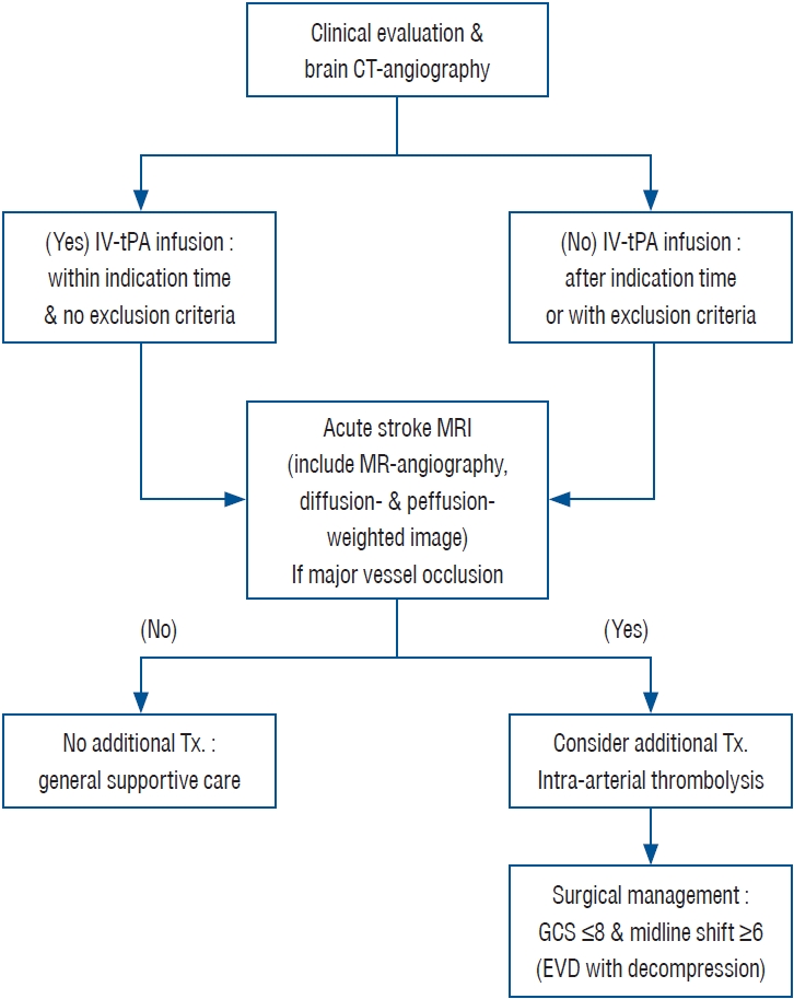
The treatment protocol was approved by the Institutional Review Board of Eunpyeong St. Mary’s Hospital (UC11RISI0187 and PC17RES10028) (Fig. 1). All patients or their representatives provided informed written consent and were made aware that they were going to receive an additional treatment after IV-tPA therapy.

Patient and neurologic examination

Between January 2010 and June 2021, 535 patients arrived within the standard treatment time from the stroke onset and were treated with IV-tPA. We excluded 235 patients because of early transfer to another hospital (16 patients), Moyamoya disease (three patients; one of these patients was transferred to another hospital), Todd’s paralysis after seizure attack (one patient), posterior circulation strokes (42 patients), tried wrong injection (one patient with subarachnoid hemorrhage) small vessel disease (126 patients). Finally, the data on anterior circulation major vessel occlusion were analyzed for 300 patients (Table 1).
All patients underwent clinical assessment (including determination of the National Institutes of Health Stroke Scale [NIHSS] score) at baseline, after 24 hours, and at 5 to 7 days, or at discharge, if earlier. A single experienced trial investigating physician, who was unaware of the treatment assignments, conducted the follow-up neurologic evaluation using the modified Rankin Scale (mRS) scale at 90±5 days when the patients visited our out-patient clinic department (Tables 1 and 2).

Neuroradiological evaluations

The neuro-radiological results were analyzed retrospectively by neuro-radiologists who had not participated in acute patient management. All patients underwent brain computed tomography angiography (CTA) (Somatom Definition AS; Siemens Medical Systems, Munich, Germany) as the initial diagnostic imaging study. If there was no flow or severe stenosis at the clinically suspected affected artery, the patient was diagnosed with LVO. Three hundred had anterior circulation LVO; their demographics are listed in Table 1. Stroke protocol magnetic resonance imaging (MRI) was performed immediately following the completion of IV-tPA administration. Obtained images included T1-weighted sagittal scans, T2-weighted turbo-gradient, spin echo/echo-planar DWI, and PWI. On the acute stroke MRI, the absence of flow signal after the suspected offending lesion as determined by the initial CTA, was correlated with non-recanalization after IV-tPA treatment. Recanalization was defined as flow that could be traced on an MRA image. Early recanalization was defined within 3 hours after IV-rtPA administration because this time window increases the chance of additional IA-Tx and is associated with better clinical outcomes [42,43].
P/D-mismatching was also evaluated on the acute stroke MRI for further analysis [9,33,47,48]. IA-Tx was recommended for non-recanalized patients with P/D-mismatching after IV-tPA treatment unless they had a malignant profile on the follow-up MRI [2]. The P/D-mismatching profile was defined as a PWI lesion that was over 100 mL and 120% or more of the diffusion lesion [27,33]. Final classification of P/D-mismatching was decided by a radiologist who was not involved in acute stroke management (Table 3). In several cases, not undertook stroke MR instead CTP and analyzed Syngovia program, not included on Table 3.
Patients who underwent IA-Tx therapy, also underwent a follow-up CT study immediately and then again within 24 hours after IA-Tx. Increased density on an immediate CT image was defined as extravasation of the contrast medium, and increased density on both follow-up CT scans was defined as a hemorrhagic complication [9,36,48]. Significant symptomatic intracranial hemorrhage was defined as neurological worsening of more than 4 points in the NIHSS score that was attributable to the presence of the clot [5,14,25].

Intra-arterial thrombectomy

Additional IA-Tx was attempted in 133 out of the 264 patients, who were not recanalized after IV-tPA treatment (Table 4). Angiograms (Axium Aristos plus; Siemens Medical Systems) were obtained using standard techniques. Once an occlusion was noted on angiography, the diagnostic catheter was exchanged for a 7-French guiding catheter (Guider Softip XF; Boston Scientific, Marlborough, MA, USA) or a balloon embolic protection device (Cello; Covidien, Irvine, CA, USA), which was placed into the internal cerebral artery. The IA-Tx was performed using stent retriever, and either Solitaire AB (EV3, Plymouth, MN, USA) or Trevo XP (Neurovascular; Stryker, Fremont, CA, USA) was selected according to the neurointerventionist’s preference.

Surgical indications for decompressive craniectomy

Additional surgical decompression was performed in 34 patients. The indications for decompressive craniectomy included the appearance of massive brain swelling on CT with clinical deterioration, worsening of the Glasgow coma scale (GCS) score below 8 and/or a midline shift of more than 6 mm and/ or obliteration of the perimesencephalic cistern on CT scans [46].If the ventricular pressure exceeded 20 mmHg after decompression surgery, conventional medical management with hyperosmotic agents, hyperventilation, and extraventricular drainage was initiated.

Statistical analyses

All data are presented as the mean±standard deviation and/or as the median. A Wilcoxon signed-rank sum test was used to analyze NIHSS scores and mRS scores. Comparisons among groups were performed using the unpaired T-test and Fisher exact test. Statistical analyses for each outcome were analyzed with SPSS software, version 20 (IBM, Armonk, NY, USA). For all statistical analyses, significance was defined by a p-value ≤0.05.

Results

Recanalization rate and treatment results after IV-tPA

The recanalization rate of LVO patients after IV-tPA was 12.0% (36/300) (Table 1). And the favorable outcome (mRS, 0-2) was 69.4% (25/36) in recanalized patients after IV-tPA treatment and 32.1% (42/131, p=0.000) in non-recanalized patients after IV-tPA treatment.
We compared the outcomes for recanalized patients (36 patients) and non-recanalized patients (131 patients) after IV-tPA without additional IA-Tx. The initial neurologic status was not different between the recanalized and non-recanalized after IV-tPA administration. The NIHSS was 12.6±6.4 (median, 11) in recanalized patients and 14.2±5.7 (median, 14) in non-recanalized patients (p=0.761) (Table 2).

Recanalization rate and treatment results after Additional IA-Tx

We attempted additional IA-Tx in 133 patients who were not recanalized by IV-tPA administration (Table 3). Additional IA-Tx was indicated in P/D-mismatching patients, based on the patients’ general condition and agreed on further additional invasive therapy. And if the patient recanalized after IA-Tx, clinical outcome was significant better (linear regression test; p=0.026).
Recanalization rate after IA-Tx was 81.1% (116/133) (Table 3). The initial neurologic status in recanalized patients was similar with non-recanalized patients (14.0±6.1 vs. 18.3±6.6, p=0.246, data not shown), but favorable outcomes were more frequent in recanalized patients than in non-recanalized patients (46.6% [54/116] vs. 22.2% [6/27], p=0.016, data not shown).

Complication rate according to P/D-mismatching

We analyzed clinical data of 167 patients treated with IV-rtPA only, and 133 treated with IA-Tx after IV-rtPA according to the P/D-mismatching or not (Table 3). In 167 patients treated IV-rtPA only, in P/D-mismatching patients (108 patients) showed more favorable clinical outcome (51/108 [47.2%] vs. 16/59 [27.1%], p=0.008), less death rate (14/108 [13.0%] vs. 21/59 [35.6%], p=0.001), more recanalization rate by IV-rtPA (29/108 [26.9%] vs. 7/59 [11.9%], p=0.018) and less clinically significant hemorrhage (8/108 [7.4%] vs. 8/59 [13.6%], p=0.042) than P/D-matching patients (Tables 3 and 4).
In 133 patients treated IA-Tx after IV-rtPA, in P/D-mismatching patients (104 patients) showed more favorable clinical outcome (52/104 [50.0%] vs. 5/29 [17.2%], p=0.001), less death rate (5/104 [4.8%] vs. 5/29 [17.2%], p=0.040), and more recanalization rate by IA-Tx (96/104 [92.3%] vs. 20/29 [69.0%], p=0.002) than P/D-matching patients. Clinically significant hemorrhage happened similarly in both groups (16/104 [15.4%] vs. 7/29 [24.1%], p=0.202), but reperfusion injury in recanalized patients (43/104 [41.3%] vs. 20/29 [69.0%], p=0.007) was less in P/D-mismatching patients (Table 3).

Discussion

Treatment for cerebral ischemic stroke is based on the ischemic penumbra [4]. According to the recanalization hypothesis, a reopening of occluded vessels before critical cell injury might improve clinical outcomes in acute ischemic stroke through regional reperfusion and salvage of threatened tissues [1,4,12,39].
As soon as intracranial bleeding is ruled out by non-contrast enhanced brain CT, application of intravenous thrombolysis for ischemic stroke is beneficial for infarction patients [20,35]. Many medical systems have been upgraded to improve clinical outcomes in infarction patients [23,38].
Recently, recanalization rate of IV-tPA LVO stroke patients has been reported [42,43]. Indeed, on these reports, the recanalization rate of LVO by OV-rtPA is very low, and has been criticized as being ineffective [7,8,10,14,16,17,19,25-27,37,41].
LVO poses a major problem and has emerged as the most common stroke subtype worldwide, especially in patients of Asian, Hispanic, and African origins [7,11,12,18,40]. The etiology and treatment of this disorder remain poorly defined. A review of previous papers suggests that medium-sized intracranial arteries and their major branches, ACA, MCA, PCA, PICA, AICA, SCA, and the distal basilar artery are most often affected [12].
The overall recanalization rate after IV-rtPA administration ranges from 10% to 47% as evaluated by transcranial sonography, CT or CTA, and other imaging techniques [3,5,7,8,10,16,17,19,23,27,32,37,39-41,45].
In our study, 535 patients were treated with IV-tPA and among them 300 patients were defined as infarction caused from LVO of anterior circulation. Considering the high incidence of LVO in our country, recanalization rate after IV-rtPA is considered for additional IA-Tx management [18]. In this study, recanalization was based on CTA and MRA, and the early recanalization rate (about 1-2 hour after IV-tPA administration) in LVO patients was 12.0% (36/300 patients).
Early recanalization is closely linked to good final clinical outcomes in acute ischemic stroke [26]. Until now, most studies performed with multimodal magnetic resonance or computed tomography imaging, the overall recanalization rate is variable about 20% up to 60%19,37,39,41). If selected patients still harbor substantial residual penumbra beyond 6 hours, they would benefit from reperfusion [2,34,39-41]. But in practice, early recanalization should be defined as recanalization within 1 hour after IV-tPA administration [3].In this study, we defined early recanalization as recanalization of the occluded larger vessel within 3 hours after IV-rtPA administration, because this time window increases chances of additional IA-Tx and is associated with better clinical outcomes [42,43].
The American Food and Drug Administration, the American Stroke Association and Korean Stroke Society have recommended IV-rtPA treatment as a standard treatment of stroke patients but after the MR CLEAN trial in 2015, IA-Tx with a stent-retrieval device became an additional treatment option after IV-rtPA [5,10,14,17,19,23,25-27,41]. The success of these studies can likely be attributed to the use of improved devices with better and faster recanalization, right patient selection with appropriate vascular imaging, and improved medical systems enabling organized care for patients treated with IA-Tx [10,17,19,25,41].
To accomplish rapid treatment, bridging therapeutic strategy, initiation of IV-tPA then followed by IA-Tx is necessary [5,8,10,14,19,25,27,41].IA-Tx enables accurate diagnosis and can facilitate mechanical clot destruction, and in some instances increases the concentration of the thrombolytic agent in the vicinity of the clot [17,47]. IA-Tx increases recanalization rate by 45.5-94% and can extend the therapeutic time window by 6-8 hours from the onset of acute ischemic stroke [10,17,19,25,41,47]. Thanks to the development of intervention devices and increased interventional experience the recanalization rate of IA-Tx is increased, and several interventionists have said that one could recanalize all the LVO cases. This aggressive approach also increased futile recanalization, significant hemorrhage rate and reperfusion injury [23,24,31,38].
Conventional non-contrast CT or MRI remains the mainstay of suspected acute stroke imaging. However, recent advanced imaging techniques, such as CTA and acute-stroke MRI, have become important tools to identify the condition of ischemic brain tissue [1,3,33]. In our study, CTA provided an initial diagnostic image, but this dynamic study did not delay IV-tPA administration. Brain radiologic evaluation, including vascular imaging, is strongly recommended in patients with severe stroke to assess large cerebral artery occlusions. In such patients, additional treatment can be prepared and save time, if IV-tPA fails to reopen the occlusion [23,27]. Neuroimaging methods for evaluating blood flow and tissue viability are increasingly required because they allow tailoring therapeutic interventions to each patient’s physiological state.
According to the authors of this study, acute stroke MRI after IV-tPA administration, especially for identifying P/D-mismatching, may identify tissue that is at risk for cerebral infarction unless blood flow is restored, and determine the risk of additional IA-Tx therapy [1,4,33].
In our study, among 133 patients who underwent IA-Tx after IV-tPA, patients with P/D-mismatching showed higher recanalization rates (92.3% in mismatching vs. 69.0% in matching, p=0.002), and higher incidence of favorable outcomes (50.0% in mismatching vs. 17.25% in matching, p=0.001). And the significant hemorrhage rate was less in P/D-mismatching patients (15.4% vs. 24.1%, p=0.202), but it was not statistically significant.
We should consider the deleterious influence of IA-Tx on hemorrhagic conversion of the infarct [36,48]. Some studies on intracerebral hemorrhage after IV-tPA reported that patient’s age, clinical stroke severity, high blood pressure, hyper-glycemia, early CT changes, and leukoaraiosis on MRI are statistically significant predictors [14,16,20,33,36,37]. Randomized studies of thrombolysis with IV-rtPA reported a significant hemorrhage rate of 1.7-8.8% [5,10,14,19,25,27,37,41].In our study, the incidence of significant hemorrhagic complications rate after IV-rtPA was 7.8%, and it was also higher in mismatching patients (7.4% vs. 13.6%, p=0.042). But in patients that undertook both IV-rtPA and IA-Tx, the significant hemorrhage was statistically similar both in P/D-mismatching and P/D-matching patients (15.4% vs. 24.1%, p=0.202), possibly because of aggressive trials to re-open the occluded vessel. And P/D-mismatching was statistically significantly correlated with favorable neurologic outcomes in IV-tPA group, IV-tPA & IA-Tx group and IA-Tx group.
Limitations of this study are that the initial imaging study was CTA and the follow-up imaging study after IV-tPA was MRI. Secondly, in our study, P/D-mismatching taken by MR imaging was analyzed to find the correlation with treatment results, but recently developed dynamic CT image (CT-perfusion) was used instead of MRI. In addition, this study is a retrospective study, and even though it was targeted to consecutive patients, some of the baseline demography-age, heart disease and atrial fibrillation-of the IA-rtPA group and the IA-Tx group showed statistical differences. More randomized, prospective studies should follow to clarify the correlation of the P/D-mismatching on MRI and dynamic CT imaging. And, the incidence of LVO is high in our country, so early recanalization rates might be different according to the reports of others.

Conclusion

If recanalization had failed after IV-tPA, additional IA-Tx might be beneficial for the patient outcome. And in this situation, P/D-mismatching on acute MR study was a good indicator for the additional IA-Tx with safety and efficacy. The authors would like to propose that we had better prepare IA-Tx when LVO is diagnosed on initial brain image. Furthermore, if the patient shows P/D-mismatching on MRA after IV-rtPA, additional IA-Tx improves treatment results and lessen the futile recanalization.

Notes

Conflicts of interest

No potential conflict of interest relevant to this article was reported.

Informed consent

Informed consent was obtained from all individual participants included in this study.

Author contributions

Conceptualization : DSY; Data curation : DSY, MHL; Formal analysis : DSY, MHL; Funding acquisition : DSY; Methodology : DSY, SHI; Project administration : DSY; Visualization : KWJ; Writing - original draft : DSY, KWJ; Writing - review & editing : DSY

Data sharing

None

Preprint

None

Acknowledgements

Authors appreciate the help of Neurosurgical nurse specialist (Min-Hee Kim, RN. & Hae-Sun Jung, RN.) to collect the data and the grammatical review of our manuscript by Ann C Rice, PhD of the J. Sargeant Reynolds Community College, Richmond, VA, USA.

Fig. 1.
Flow diagram of treatment protocol. CTA : computed tomography angiography, IV-tPA : intravenous tissue plasminogen activator administration, MRI : magnetic resonance imaging, Tx. : treatment, GCS : Glasgow coma scale, EVD : extraventriular drainage.
jkns-2022-0120f1.jpg
Table 1.
Demography and clinical outcomes of the 300 patients with anterior circulation LVO, according to the treatments
IV-tPA only IV-tPA & IA-Tx Total statistic value p-value
Patients No. 167 133 300
Age (years) 67.4±11.5 64.0±15.0 65.9±13.3 0.046
 Median 69 (39-89) 65 (19-98) 68 (19-98)
Male sex 101 (60.5) 88 (66.2) 189 (63.0) 0.186
NIHSS score 13.9±5.9 14.7±6.4 14.2±6.1 0.284
 Median 13 (4-32) 14 (4-36) 14 (4-36)
Heart disease 38 (29.9) 16 (12.0) 54 (18.1) 0.011
Atrial fibrillation 72 (43.1) 42 (31.6) 114 (38.0) 0.027
Hypertension 99 (59.3) 74 (55.6) 173 (57.7) 0.303
Dyslipidemia 49 (32.9) 31 (23.8) 80 (28.7) 0.062
Diabetic mellitus 50 (30.1) 43 (32.3) 93 (31.1) 0.387
Smoking 42 (25.3) 31 (23.7) 73 (24.6) 0.426
Occlusion vessel site
 ACA 5 (3.0) 5 (1.7)
 MCA Lt. 45 (26.9) 36 (27.1) 81 (27.0)
 MCA Rt. 66 (39.5) 49 (36.8) 115 (38.3)
 ICA Lt. 27 (16.2) 23 (17.3) 50 (16.7)
 ICA Rt. 24 (14.4) 25 (18.8) 49 (16.3)
Time to tPA (minutes) 128.4±51.5 123.6±55.8 126.3±53.4
 Median 119 (45-270) 110 (45-270) 120 (45-270) 0.214
Time to femoral artery puncture (hours) 5.4±1.7 (5.0)
 Median 5 (1-11)
Recanalized after IV-tPA 36 (12.0)
Recanalized after IA-Tx 113 (87.3)
Neurologic outcomes
 mRS
  0 23 (13.8) 19 (14.3) 42 (14.0)
  1 22 (13.2) 19 (14.3) 41 (13.7)
  2 22 (13.2) 21 (15.8) 43 (14.3)
 Favorable 67 (40.1) 59 (44.4) 126 (41.3) 0.068
  3 35 (21.0) 27 (20.3) 62 (20.7)
  4 14 (8.4) 21 (15.8) 35 (11.7)
  5 16 (9.6) 16 (12.0) 32 (10.7)
 Unfavorable 65 (38.9) 64 (49.6) 129 (43.7)
  6 35 (21.0) 10 (7.5) 45 (15.0) 0.001
Sx-Hx 13 (7.8) 22 (16.5) 35 (11.7) 0.010
Reperfusion injury 17 (13.0) 63 (47.4) 86 (28.7) <0.001
Decompressive surgery 18 (10.8) 16 (12.0) 34 (11.3) 0.436
P/D-mismatching 108 (64.7) 104 (78.2) 212 (70.7) 0.008

Values are presented as mean±standard deviation, number (range), or number (%). LVO : large vessel occlusion, IV-tPA : intravenous tissue plasminogen activator administration, IA-Tx : intra-arterial thrombolytic therapy, NIHSS : National Institutes of Health Stroke Scale, ACA : anterior cerebral artery, MCA : middle cerebral artery, Lt. : left, Rt. : right, ICA : internal cerebral artery, mRS : modified Rankin Scale, Sx-Hx : symptomatic hemorrhage, P/D : perfusion/diffusion

Table 2.
Clinical characteristic and outcomes of the IV-rtPA treated, 167 anterior circulation large vessel occlusion patients
Recanal after IV-tPA Not recanal after IV-tPA Total statistic value p-value
Patients No. 36 (12.0) 131 (78.4) 167
Age (years) 64.9±11.6 68.1±11.4 67.4±11.5 0.766
 Median 68 (42-82) 70 (39-89) 69 (39-89)
Male sex 24 (66.7) 77 (58.8) 101 (60.5) 0.255
NIHSS score 12.6±6.4 14.2±5.7 13.9±5.9 0.761
 Median 11 (4-32) 14 (4-32) 13 (4-32)
Heart disease 4 (11.1) 34 (26.2) 38 (22.9) 0.041
Atrial fibrillation 13 (36.1) 59 (45.0) 72 (43.1) 0.222
Hypertension 15 (41.7) 84 (64.1) 99 (59.3) 0.013
Dyslipidemia 11 (35.5) 38 (32.2) 49 (32.9) 0.442
Diabetic mellitus 11 (30.6) 39 (30.0) 50 (30.1) 0.550
Smoking 8 (22.2) 34 (26.2) 42 (25.3) 0.404
Alcohol 11 (30.6) 39 (30.7) 50 (30.7) 0.580
Occlusion vessel site
 ACA 0 (0.0) 5 (3.8) 5 (3.0)
 MCA Lt. 11 (30.6) 34 (26.0) 45 (26.9)
 MCA Rt. 23 (63.9) 43 (32.8) 66 (39.5)
 ICA Lt. 0 (0.0) 27 (20.6) 27 (16.2)
 ICA Rt. 2 (5.6) 22 (16.8) 24 (14.4)
Time to tPA (minutes) 121.7±57.0 130.0±50.0 128.4±51.5 0.619
 Median 120 (45-270) 120 (45-270) 119 (45-270)
Neurologic outcomes
 mRS
  0 12 (33.3) 11 (8.4) 23 (13.8)
  1 8 (22.2) 14 (10.7) 22 (13.2)
  2 5 (13.9) 17 (13.0) 22 (13.2)
 Favorable 25 (69.4) 42 (32.1) 67 (40.1) <0.001
  3 6 (16.7) 29 (22.1) 35 (21.0)
  4 1 (2.8) 13 (9.9) 14 (8.4)
  5 16 (12.2) 16 (9.6)
 Unfavorable 7 (19.5) 58 (44.3) 65 (38.9)
  6 4 (11.1) 31 (23.7) 35 (21.0) 0.075
Sx-Hx 3 (8.3) 10 (7.6) 13 (7.8) 0.561
Decompressive surgery 2 (5.6) 16 (12.2) 18 (10.8) 0.206
P/D-mismatching 29 (80.6) 79 (60.3) 108 (64.7) 0.018

Values are presented as mean±standard deviation, number (range), or number (%). IV-rtPA : intravenous recombinant tissue plasminogen activator, IV-tPA : intravenous tissue plasminogen activator administration, IA-Tx : intra-arterial thrombolytic therapy, NIHSS : National Institutes of Health Stroke Scale, ACA : anterior cerebral artery, MCA : middle cerebral artery, Lt. : left, Rt. : right, ICA : internal cerebral artery, mRS : modified Rankin Scale, Sx-Hx : symptomatic hemorrhage, P/D : perfusion/diffusion

Table 3.
Patient characteristics and clinical outcomes according to the treatment methods and P/D-mismatching or not
IV-tPA (n=167)
IV-tPA & IA-Tx (n=133)
Mismatched Matched p-value Mismatched Matched p-value
Patients No. 108 59 104 29
Age (years) 67.4±11.8 67.7±11.0 0.827 64.6±15.4 61.8±13.4 0.538
 Median 69 (39-89) 69 (46-86) 65 (19-98) 62 (24-82)
Male sex 62 (57.4) 39 (66.1) 0.176 66 (63.5) 22 (75.9) 0.152
NIHSS score 12.9±5.1 15.8±6.8 0.006 13.8±6.0 18.0±6.7 0.685
 Median 13 (4-28) 15 (4-32) 13 (4-36) 17 (5-32)
Time to tPA (minutes) 126.5±50.0 131.9±55.1 0.557 123.5±54.6 130.2±53.7 0.738
 Median 120 (45-270) 120 (48-270) 110 (45-270) 116 (65-270)
Femoral artery puncture time (hours) 5.0±2.1 5.2±1.6 0.312
 Median 50 (1.5-15) 50 (2.5-11)
Recanalized by IV-rtPA or IA-Tx 29 (26.9) 7 (11.9) 0.018 96 (92.3) 20 (69.0) 0.002
Neurologic outcomes
 Favorable 51 (47.2) 16 (27.1) 0.008 52 (50.0) 5 (17.2) 0.001
 Unfavorable 43 (39.8) 22 (37.3) 19 (45.2) 19 (65.6)
 Dead 14 (13.0) 21 (35.6) 0.001 5 (4.8) 5 (17.2) 0.040
Sx-Hx 8 (7.4) 8 (13.6) 0.042 16 (15.4) 7 (24.1) 0.202
Reperfusion injury 13 (12.0) 10 (16.9) 0.256 43 (41.3) 20 (69.0) 0.007
Decompressive surgery 9 (8.3) 9 (15.3) 0.133 7 (6.7) 9 (31.0) 0.001

Values are presented as mean±standard deviation, number (range), or number (%). P/D : perfusion/diffusion, IV-tPA : intravenous tissue plasminogen activator administration, IA-Tx : intra-arterial thrombolytic therapy, NIHSS : National Institutes of Health Stroke Scale, Sx-Hx : symptomatic hemorrhage

Table 4.
Two hundred sixty-four patient clinical results after IA-Tx, who were not recanalized by IV-rtPA
IV-rtPA without IA-Tx IV-tPA with add IA-Tx Total statistic value p-value
Patients No. 131 133 264
Age (years) 68.1±11.4 64.0±15.0 66.0±13.5 0.056
 Median 70 (39-89) 65 (19-98) 68 (19-98)
Male sex 77 (58.8) 88 (66.2) 165 (62.5) 0.133
NIHSS score 14.2±5.8 14.7±6.4 14.5±6.1 0.199
 Median 14 (4-32) 14 (4-36) 14 (4-36)
Occlusion vessel site
 ACA 5 (3.8) 5 (1.9)
 MCA Lt. 34 (26.0) 36 (27.1) 70 (26.5)
 MCA Rt. 43 (32.8) 49 (36.8) 92 (34.8)
 ICA Lt. 27 (20.6) 23 (17.3) 50 (18.9)
 ICA Rt. 22 (16.8) 27 (18.8) 47 (17.8)
Time to tPA (minutes) 121.7±57.0 123.6±55.8 127.6±52.5 0.191
 Median 120 (51-270) 100 (45-270) 120 (45-270)
Time to femoral artery puncture (hours) 5.0±1.8 5.0±1.8
 Median 5.0 (1.5-11.0) 5.0 (1.5-11.0)
Recanalization after IA-Tx 116 (87.3)
Neurologic outcomes
 mRS
  0 11 (8.4) 19 (14.3) 30 (11.4)
  1 14 (10.7) 19 (14.3) 33 (12.5)
  2 17 (13.0) 21 (15.8) 38 (14.4)
 Favorable 42 (32.1) 57 (42.9) 99 (37.5) 0.046
  3 29 (22.1) 27 (20.3) 56 (21.2)
  4 13 (9.9) 21 (15.3) 34 (12.9)
  5 16 (12.2) 16 (12.0) 32 (12.1)
 Unfavorable 58 (44.3) 66 (49.6) 124 (47.0)
  6 31 (23.7) 10 (7.5) 41 (15.5) <0.001
Sx-Hx 10 (7.6) 22 (16.5) 33 (12.5) 0.014
Decompressive surgery 16 (12.2) 16 (12.0) 32 (12.1) 0.557
P/D-mismatching 79 (60.3) 104 (78.2) 183 (69.3) 0.001

Values are presented as mean±standard deviation, number (range), or number (%). IA-Tx : intra-arterial thrombolytic therapy, IV-rtPA : intravenous recombinant tissue plasminogen activator, IV-tPA : intravenous tissue plasminogen activator administration, NIHSS : National Institutes of Health Stroke Scale, ACA : anterior cerebral artery, MCA : middle cerebral artery, Lt. : left, Rt. : right, ICA : internal cerebral artery, mRS : modified Rankin Scale, Sx-Hx : symptomatic hemorrhage, P/D : perfusion/diffusion

References

1. Abe O, Aoki S, Shirouzu I, Kunimatsu A, Hayashi N, Masumoto T, et al : MR imaging of ischemic penumbra. Eur J Radiol 46 : 67-78, 2003
crossref pmid
2. Albers GW, Thijs VN, Wechsler L, Kemp S, Schlaug G, Skalabrin E, et al : Magnetic resonance imaging profiles predict clinical response to early reperfusion: the diffusion and perfusion imaging evaluation for understanding stroke evolution (DEFUSE) study. Ann Neurol 60 : 508-517, 2006
crossref pmid pdf
3. Aoki J, Kimura K, Shibazaki K, Sakamoto Y : DWI-ASPECTS as a predictor of dramatic recovery after intravenous recombinant tissue plasminogen activator administration in patients with middle cerebral artery occlusion. Stroke 44 : 534-537, 2013
crossref pmid
4. Astrup J, Siesjö BK, Symon L : Thresholds in cerebral ischemia - the ischemic penumbra. Stroke 12 : 723-725, 1981
crossref pmid
5. Berkhemer OA, Fransen PS, Beumer D, van den Berg LA, Lingsma HF, Yoo AJ, et al : A randomized trial of intraarterial treatment for acute ischemic stroke. N Engl J Med 372 : 11-20, 2015
pmid
6. Berrouschot J, Sterker M, Bettin S, Köster J, Schneider D : Mortality of space-occupying (‘malignant’) middle cerebral artery infarction under conservative intensive care. Intensive Care Med 24 : 620-623, 1998
crossref pmid pdf
7. Bhatia R, Hill MD, Shobha N, Menon B, Bal S, Kochar P, et al : Low rates of acute recanalization with intravenous recombinant tissue plasminogen activator in ischemic stroke: real-world experience and a call for action. Stroke 41 : 2254-2258, 2010
crossref pmid
8. Broderick JP, Berkhemer OA, Palesch YY, Dippel DW, Foster LD, Roos YB, et al : Endovascular therapy is effective and safe for patients with severe ischemic stroke: pooled analysis of interventional management of stroke III and multicenter randomized clinical trial of endovascular therapy for acute ischemic stroke in the Netherlands data. Stroke 46 : 3416-3422, 2015
crossref pmid pmc
9. Butcher KS, Parsons M, MacGregor L, Barber PA, Chalk J, Bladin C, et al : Refining the perfusion-diffusion mismatch hypothesis. Stroke 36 : 1153-1159, 2005
crossref pmid
10. Campbell BC, Mitchell PJ, Kleinig TJ, Dewey HM, Churilov L, Yassi N, et al : Endovascular therapy for ischemic stroke with perfusion-imaging selection. N Engl J Med 372 : 1009-1018, 2015
crossref pmid
11. Caplan LR : Intracranial large artery occlusive disease. Curr Neurol Neurosci Rep 8 : 177-181, 2008
crossref pmid pdf
12. Caplan LR, Gorelick PB, Hier DB : Race, sex and occlusive cerebrovascular disease: a review. Stroke 17 : 648-655, 1986
crossref pmid
13. Chimowitz MI, Lynn MJ, Howlett-Smith H, Stern BJ, Hertzberg VS, Frankel MR, et al : Comparison of warfarin and aspirin for symptomatic intracranial arterial stenosis. N Engl J Med 352 : 1305-1316, 2005
crossref pmid
14. Ciccone A, Valvassori L, Nichelatti M, Sgoifo A, Ponzio M, Sterzi R, et al : Endovascular treatment for acute ischemic stroke. N Engl J Med 368 : 904-913, 2013
crossref pmid pmc
15. Davis S, Donnan GA : Time is penumbra: imaging, selection and outcome. the Johann Jacob Wepfer award 2014. Cerebrovasc Dis 38 : 59-72, 2014
crossref pmid pdf
16. De Silva DA, Brekenfeld C, Ebinger M, Christensen S, Barber PA, Butcher KS, et al : The benefits of intravenous thrombolysis relate to the site of baseline arterial occlusion in the Echoplanar Imaging Thrombolytic Evaluation Trial (EPITHET). Stroke 41 : 295-299, 2010
crossref pmid
17. Furlan A, Higashida R, Wechsler L, Gent M, Rowley H, Kase C, et al : Intra-arterial prourokinase for acute ischemic stroke. the PROACT II study: a randomized controlled trial. prolyse in acute cerebral thromboembolism. JAMA 282 : 2003-2011, 1999
crossref pmid
18. Gorelick PB, Wong KS, Bae HJ, Pandey DK : Large artery intracranial occlusive disease: a large worldwide burden but a relatively neglected frontier. Stroke 39 : 2396-2399, 2008
crossref pmid
19. Goyal M, Demchuk AM, Menon BK, Eesa M, Rempel JL, Thornton J, et al : Randomized assessment of rapid endovascular treatment of ischemic stroke. N Engl J Med 372 : 1019-1030, 2015
crossref pmid
20. Hacke W, Kaste M, Bluhmki E, Brozman M, Dávalos A, Guidetti D, et al : Thrombolysis with alteplase 3 to 4.5 hours after acute ischemic stroke. N Engl J Med 359 : 1317-1329, 2008
crossref pmid
21. Hacke W, Schwab S, Horn M, Spranger M, De Georgia M, von Kummer R : ‘Malignant’ middle cerebral artery territory infarction: clinical course and prognostic signs. Arch Neurol 53 : 309-315, 1996
crossref pmid
22. Hofmeijer J, Kappelle LJ, Algra A, Amelink GJ, van Gijn J, van der Worp HB, et al : Surgical decompression for space-occupying cerebral infarction (the Hemicraniectomy After Middle Cerebral Artery infarction with Life-threatening Edema Trial [HAMLET]): a multicentre, open, randomised trial. Lancet Neurol 8 : 326-333, 2009
crossref pmid
23. Hong KS, Ko SB, Yu KH, Jung C, Park SQ, Kim BM, et al : Update of the Korean clinical practice guidelines for endovascular recanalization therapy in patients with acute ischemic stroke. J Stroke 18 : 102-113, 2016
crossref pmid pmc pdf
24. Hutchinson PJ, Kolias AG, Timofeev IS, Corteen EA, Czosnyka M, Timothy J, et al : Trial of decompressive craniectomy for traumatic intracranial hypertension. N Engl J Med 375 : 1119-1130, 2016
crossref pmid
25. Jovin TG, Chamorro A, Cobo E, de Miquel MA, Molina CA, Rovira A, et al : Thrombectomy within 8 hours after symptom onset in ischemic stroke. N Engl J Med 372 : 2296-2306, 2015
crossref pmid
26. Khatri P, Abruzzo T, Yeatts SD, Nichols C, Broderick JP, Tomsick TA, et al : Good clinical outcome after ischemic stroke with successful revascularization is time-dependent. Neurology 73 : 1066-1072, 2009
crossref pmid pmc
27. Kidwell CS, Jahan R, Gornbein J, Alger JR, Nenov V, Ajani Z, et al : A trial of imaging selection and endovascular treatment for ischemic stroke. N Engl J Med 368 : 914-923, 2013
crossref pmid pmc
28. Larrue V, von Kummer RR, Müller A, Bluhmki E : Risk factors for severe hemorrhagic transformation in ischemic stroke patients treated with recombinant tissue plasminogen activator: a secondary analysis of the European-Australasian Acute Stroke Study (ECASS II). Stroke 32 : 438-441, 2001
crossref pmid
29. Lewandowski CA, Frankel M, Tomsick TA, Broderick J, Frey J, Clark W, et al : Combined intravenous and intra-arterial r-TPA versus intra-arterial therapy of acute ischemic stroke: Emergency Management of Stroke (EMS) bridging trial. Stroke 30 : 2598-2605, 1999
crossref pmid
30. Lin J, Frontera JA : Decompressive hemicraniectomy for large hemispheric strokes. Stroke 52 : 1500-1510, 2021
crossref pmid
31. Mazighi M, Serfaty JM, Labreuche J, Laissy JP, Meseguer E, Lavallée PC, et al : Comparison of intravenous alteplase with a combined intravenous-endovascular approach in patients with stroke and confirmed arterial occlusion (RECANALISE study): a prospective cohort study. Lancet Neurol 8 : 802-809, 2009
crossref pmid
32. Mishra SM, Dykeman J, Sajobi TT, Trivedi A, Almekhlafi M, Sohn SI, et al : Early reperfusion rates with IV tPA are determined by CTA clot characteristics. AJNR Am J Neuroradiol 35 : 2265-2272, 2014
crossref pmid pmc
33. Mlynash M, Lansberg MG, De Silva DA, Lee J, Christensen S, Straka M, et al : Refining the definition of the malignant profile: insights from the DEFUSE-EPITHET pooled data set. Stroke 42 : 1270-1275, 2011
crossref pmid pmc
34. Muchada M, Rodriguez-Luna D, Pagola J, Flores A, Sanjuan E, Meler P, et al : Impact of time to treatment on tissue-type plasminogen activator-induced recanalization in acute ischemic stroke. Stroke 45 : 2734-2738, 2014
crossref pmid
35. National Institute of Neurological Disorders and Stroke rt-PA Stroke Study Group : Tissue plasminogen activator for acute ischemic stroke. N Engl J Med 333 : 1581-1587, 1995
crossref pmid
36. Paciaroni M, Agnelli G, Corea F, Ageno W, Alberti A, Lanari A, et al : Early hemorrhagic transformation of brain infarction: rate, predictive factors, and influence on clinical outcome: results of a prospective multicenter study. Stroke 39 : 2249-2256, 2008
crossref pmid
37. Paciaroni M, Balucani C, Agnelli G, Caso V, Silvestrelli G, Grotta JC, et al : Systemic thrombolysis in patients with acute ischemic stroke and Internal Carotid ARtery Occlusion: the ICARO study. Stroke 43 : 125-130, 2012
crossref pmid
38. Powers WJ, Rabinstein AA, Ackerson T, Adeoye OM, Bambakidis NC, Becker K, et al : 2018 guidelines for the early management of patients with acute ischemic stroke: a guideline for healthcare professionals from the American Heart Association/American Stroke Association. Stroke 49 : e46-e110, 2018
crossref pmid
39. Rha JH, Saver JL : The impact of recanalization on ischemic stroke outcome: a meta-analysis. Stroke 38 : 967-973, 2007
crossref pmid
40. Ribo M, Alvarez-Sabín J, Montaner J, Romero F, Delgado P, Rubiera M, et al : Temporal profile of recanalization after intravenous tissue plasminogen activator: selecting patients for rescue reperfusion techniques. Stroke 37 : 1000-1004, 2006
crossref pmid
41. Saver JL, Goyal M, Bonafe A, Diener HC, Levy EI, Pereira VM, et al : Stent-retriever thrombectomy after intravenous t-PA vs. t-PA alone in stroke. N Engl J Med 372 : 2285-2295, 2015
crossref pmid
42. Seners P, Turc G, Maïer B, Mas JL, Oppenheim C, Baron JC : Incidence and predictors of early recanalization after intravenous thrombolysis: a systematic review and meta-analysis. Stroke 47 : 2409-2412, 2016
crossref pmid
43. Tsivgoulis G, Katsanos AH, Schellinger PD, Köhrmann M, Varelas P, Magoufis G, et al : Successful reperfusion with intravenous thrombolysis preceding mechanical thrombectomy in large-vessel occlusions. Stroke 49 : 232-235, 2018
crossref pmid pmc
44. Wardlaw JM, Murray V, Berge E, del Zoppo G, Sandercock P, Lindley RL, et al : Recombinant tissue plasminogen activator for acute ischaemic stroke: an updated systematic review and meta-analysis. Lancet 379 : 2364-2372, 2012
crossref pmid pmc
45. Wei XE, Zhao YW, Lu J, Li MH, Li WB, Zhou YJ, et al : Timing of recanalization and outcome in ischemic-stroke patients treated with recombinant tissue plasminogen activator. Acta Radiol 56 : 1119-1126, 2015
crossref pmid pdf
46. Yoo DS, Kim DS, Cho KS, Huh PW, Park CK, Kang JK : Ventricular pressure monitoring during bilateral decompression with dural expansion. J Neurosurg 91 : 953-959, 1999
crossref pmid
47. Yoo DS, Won YD, Huh PW, Shin HE, Kim KT, Kang SG, et al : Therapeutic results of intra-arterial thrombolysis after full-dose intravenous tissue plasminogen activator administration. AJNR Am J Neuroradiol 31 : 1536-1540, 2010
crossref pmid pmc
48. Yoon W, Seo JJ, Kim JK, Cho KH, Park JG, Kang HK : Contrast enhancement and contrast extravasation on computed tomography after intraarterial thrombolysis in patients with acute ischemic stroke. Stroke 35 : 876-881, 2004
crossref pmid
TOOLS
PDF Links  PDF Links
PubReader  PubReader
ePub Link  ePub Link
XML Download  XML Download
Full text via DOI  Full text via DOI
Download Citation  Download Citation
  Print
Share:      
METRICS
1
Crossref
1
Scopus
2,846
View
116
Download
Related article
Editorial Office
1F, 18, Heolleung-ro 569-gil, Gangnam-gu, Seoul, Republic of Korea
TEL: +82-2-525-7552   FAX: +82-2-525-7554   E-mail: office@jkns.or.kr
About |  Browse Articles |  Current Issue |  For Authors and Reviewers
Copyright © Korean Neurosurgical Society.                 Developed in M2PI
Close layer