INTRODUCTION
Traumatic spinal cord injury (SCI) resulting in motor weakness, sensory loss, and autonomic dysfunction, has no efficacious clinical treatments. In past years, researchers have sought to find efficacious therapies for the treatment of traumatic SCI [1,6-8,10,11,14], many of which have been found to be effective in in vivo rodent models of SCI. Unfortunately, while many experimental treatments have shown great promise in rodent models, the few of them that have actually successfully been translated into clinical trials for human SCI, have failed to demonstrate convincing efficacy [24]. While rodent models of SCI have many benefits, including well-controlled injuries (contusion impactors, clip compression, dorsal hemisection), and widely utilized outcome measures (Basso, Beattie, Bresnahan locomotor scale, cylinder rearing test, horizontal ladder test, catwalk), the anatomical differences in cord size, and subarachnoid space compared with humans are notable (Fig. 1). These differences may contribute to the lack of success of clinical trials for SCI therapies, which otherwise show promise in small animal, rodent studies.
Here, we review the large animal model of SCI using Yucatan miniature pigs by Lee et al. [15]. Although there are challenges with regards to experimental cost and animal housing of larger species, the pig model has a number of advantages as a preclinical model of traumatic SCI. The anatomical, physiological, and genetic similarities between pigs [27] and humans have made pigs useful as models in a variety of research settings including stroke [3,9,17], traumatic brain injury [2,25,28], and SCI [18,21-23,26]. We suggest that the porcine model of thoracic SCI is a useful intermediary model for preclinical SCI studies. In the present review, we describe our porcine model of SCI and present examples of how this model can be used for studying the pathophysiology of traumatic SCI as well as validating results from promising rodent studies.
SURGICAL PROCEDURES
The description of the development of our pig model was published by Lee et al. [15] with details of the surgical procedures.
Anesthesia and preparation for surgery
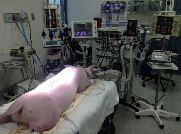
Our model utilizes female Yucatan miniature pigs typically weighing between 20-25 kg at the time of surgery. Animals are pre-medicated with telazol 4-6 mg/kg intramuscular (IM) injection in conjunction with xylazine 1 mg/kg IM and atropine (0.02-0.04 mg/kg) IM. Animals are induced with either isoflurane (2-3% in O2) or propofol 2 mg/kg, depending on the studyŌĆÖs anesthetic requirements, and endotracheally intubated. Pigs are given an intravenous (IV) dose of antibiotics (cefazolin, 15 mg/kg), and an IM injection of analgesics (ketoprofen, 3 mg/kg) before surgery. Bupivacaine (1 mg/kg) is injected into the skin and subcutaneous tissue of the incision line as a local anesthetic. A urinary catheter (10 Fr, Jorgensen Laboratories Inc., Loveland, CO, USA) is manually inserted before surgery and is maintained after surgery until the spontaneous urination is restored. Mechanical ventilation is maintained at 10-12 breaths/min and the tidal volume at 12-15 mL/kg (Veterinary Anesthesia Ventilator model 2002, Hallowell EMC, Pittsfield, MA, USA). Standard monitoring is performed for heart rate, respiratory rate, blood pressure, endtidal carbon dioxide, inspired and expired isoflurane levels, and oxygen saturation (pulse oximeter 8600V, Nonin Medical Inc., Markham, ON, USA and Cardell MAX-12HD Veterinary Monitor, Paragon Medical Supply, Inc., Coral Springs, FL, USA). Hydration is maintained with 1.25% dextrose solution and the temperature is measured by a rectal probe and maintained at 37.0-38.0oC with a heating pad (T/Pump, Gaymar Industries, Inc., Orchard Park, NY, USA).
Laminectomy and mounting of platform
Our most commonly performed injury is at the level of Thoracic 10 (T10) level, but the injury level can be altered depending on the research design. The vertebral levels are easily identified with anatomical landmarks such as the apex of the kyphotic thoracic spine. With the pig in the prone position on the surgical table (Fig. 2), a dorsal midline incision is made between T7 and T14, the length of which is determined by the nature of the experiment. The spinous processes, laminae, and transverse processes of T8-T13 are exposed using monopolar electrocautery. The transverse processes are then removed using a rongeur in order to place pedicle screws at the T6 and T8 or T11 and T13 levels, using 3.5├Ś25 mm poly-axial screws (Vertex screws, Medtronic, Memphis, TN, USA) that are designed for human cervical spine fixation. The location of screw insertion can vary depending on the experimental design. A titanium rod is fixed with the locking caps to mount the weight drop guidance system, to rigidly fix the T9-12 segments during the weight drop, and to provide stability to the spine after SCI. A laminectomy is performed at the T8-T11 level and widened to ensure that a circular window measuring at least 1.2 cm in diameter (impactor diameter, 0.95 cm) is made to expose the dura and spinal cord.
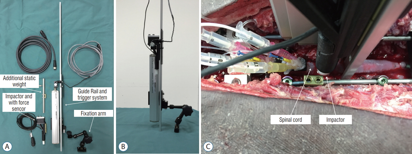
SCI
Five minutes before SCI, isof lurane is increased to 3.0% from 0.5% to minimize involuntary movement as a result of the SCI. The weight drop apparatus consists of a guidance rail along which a 50 g impactor falls onto the exposed spinal cord from a defined height (Fig. 3) [5,15]. The guidance system for the impactor is secured rigidly to the rods held by the pedicle screws, and by adjusting the articulating arm, it is positioned directly over the laminectomy defect. The rail guidance system is leveled to ensure that it is aligned vertically in both the coronal and sagittal planes. The laminectomy defect size is checked to ensure that the impactor tip can fall freely on the midline of the dura and spinal cord without any interference. A load cell (LLB215, Futek Advanced Sensor Technology, Irvine, CA, USA) is installed at the end of the impact device to record the force during impact. Finally, the impactor is released and falls along the guidance rail system, and impacts the spinal cord. Immediately after the contusion injury, we placed a 100 g weight on top of the 50 g weight impactor to simulate sustained compression (total 150 g). The duration of compression can be adjusted depending upon the experiment - typically we use 5 minutes of sustained compression. Following 5 minutes of sustained compression, the weight drop apparatus is removed and the wound is closed by layer. However, we could control the degree of cord damage by the impactor height, weight, and the additional compression duration. In the previous study, we analyzed the effect of impactor height on tissue damage by dropping it from various heights - 50, 40, 30, 20, and 10 cm [15]. The height of the impactor drop was very well correlated with the histologic and function outcomes. Also, the additional compression duration and weight after primary impact could be another important factor in SCI, because the contusion from a height of 20 cm with or without sustained compression showed significant metabolic and histologic difference [18]. We ultimately chose 20 cm for the height of the impactor drop, combined with 5 minutes of compression, which produces a lower extremity paralysis that recovers to a Porcine Thoracic Injury Behavioral Scale (PTIBS) of approximately 4 at 12 weeks post-injury (described below).
After the surgery, the animal is maintained on a continuous rate infusion of fentanyl for pain control, which requires close observation. The animals are individually housed in recovery kennels for 7 days. The urinary catheter is removed 7-10 days after SCI, once the animals are able to reflexively empty their bladders. Animals are then housed in pairs on sawdust bedding for 12 weeks, during which behavioral recovery analysis is performed weekly.
PARAMETERS ANALYSIS
Biomechanical analysis
The data acquired from the impactor tip during the actual contusive injury is collected using a custom Labview (V8.6, National Instruments, Austin, TX, USA) program at 100 kHz, followed by post-filtering and then customized with Matlab (V2008b, The Mathworks, Matick, MA, USA) using a bi-directional 4-way filter with 5 kHz low-pass cutoff frequency. Injury parameters such as impact force, displacement, velocity, and impulse are measured, providing relevant information on injury severity and variability.
Functional outcome - the PTIBS
To characterize hindlimb locomotor recovery, we created the Porcine Thoracic Injury Behavior Scale (PTIBS) [15]. The PTIBS stratifies hind limb locomotor function into ten categories, ranging from absent hind limb movement (score 1) to normal walking (score 10). A PTIBS score of 1-3 ref lects ŌĆ£hindlimb draggingŌĆØ, scores of 4-6 reflect varying degrees of ŌĆ£steppingŌĆØ ability, and scores of 7-10 reflect varying degrees of ŌĆ£walkingŌĆØ ability. A detailed description and functional components of each PTIBS score are listed in Tables 1 and 2, respectively. Hindlimb function is recorded using three highdefinition camcorders located behind the animal, so that the observer can visualize the movement of the hindlimbs and hips as the animal moves away from the camera. One week before the injury, videos are recorded of the animals to establish a ŌĆ£baselineŌĆØ locomotor behavior score. If an animal fails to achieve a score of 10 at baseline, the animal will be removed from the study. Post-injury, videos are recorded once per week for 12 weeks and subsequently scored by two independent, blinded observers.
The reliability of PTIBS is relatively high under conditions of proper training. In our previous study [15], 154 video clips were scored by three observers (two trained technicians and an undergraduate student) on two occasions, 3 weeks apart. The intra-observer correlation coefficients were 0.954, 0.992, and 0.972 for the three observers and the inter-observer correlation coefficients were 0.961, 0.927, and 0.918. In addition, we analyzed the relationship between the histologic damage of the spinal cord and PTIBS. The PTIBS scores are positively correlated with white and gray matter sparing (see below in the ŌĆ£Histologic analysisŌĆØ) 12 weeks post-injury. The correlation coefficient between the PTIBS score and the spared white matter was R=0.888 (p <0.001) and gray matter was q=0.748 (p <0.001). Also, the PTIBS scores are highly correlated with total (white and gray matter) spared tissue with a coefficient of q=0.881 (p <0.001). Also, the neurofilament immunoreactivity showed a positive correlation with the PTIBS score as a coefficient of q=0.663 (p =0.002).
In a recent study [26], we analyzed the correlation between the impact degree and PTIBS. The impact degree falls into three groups; mild, moderate, and severe. A mild impact degree represents an injury from a 10 cm drop height, moderate represents 20 cm, and severe represents 40 cm. One week after SCI, functional outcomes were most severely impaired. The average PTIBS score for the mild, moderate, and severe injury groups, were 4.0, 3.2, and 2.0, respectively. At 12 weeks after injury, the average PTIBS scores were 6.5, 4.25, and 3.0, respectively. Impact height had a significant effect on the PTIBS scores (p<0.0001) between all injury groups from 6 to 12 weeks after SCI. This data suggests that the impact force is strongly correlated with behavioral recovery.
Histologic analysis
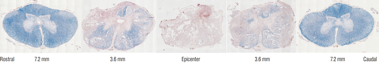
For histological evaluation of damage induced by injury, a 10 cm segment of the thoracic spinal cord centered around the lesion is collected, post-fixed in 4% paraformaldehyde for 86 hours at 4oC, and cryopreserved by transferring the tissue through a series of solutions with gradually increasing sucrose concentrations (12%, 22%, and 30%). Spinal cord segments are divided into 1 cm blocks, one of them centering the impact site, kept frozen at -80oC, and cryo-sectioned with a thickness of 20 ╬╝m.
For the measurement of spared white and gray matter, a series of cross-sections are stained with Eriochrome Cyanine R as described in Rabchevsky et al. [19] and counterstained with Neutral Red (Fig. 4). The images are then captured with an optical microscope (Leica DM5000B, Leica, Concord, Canada) using a 2.5├Ś or 5.0├Ś objective lens. In the uninjured tissue, the staining allows clear differentiation between the white matter, gray matter and the pia by the color of the stain and the shape. In the injured tissue, the spared white matter is defined as a region showing dense blue staining. We defined uninjured gray matter as tissue containing a normal gray matter cytoarchitecture and keeping the residual blue color with visible neutral red staining speck. The cumulative preserved white and gray matter is calculated by summing the percentage of spared white and gray matter in a segment around epicenter, for example from 1.52 cm rostral to 1.52 cm caudal to the epicenter [16]. This provides a general volumetric representation of the extent of the injury.
In a recent study [26], we analyzed the correlation between the impact degree (sham, mild, moderate, severe) and histology. The height of injury had a significant effect on the amount of spared tissue, calculated with the area under the curve of the total percent spared tissue from 13.6 mm rostral and 13.6 mm caudal of the epicenter (p <0.0001). Cumulative spared tissue was greatest in the SHAM group (3380┬▒16.0 cumulative%) compared to the mild (2720┬▒98.0 cumulative%, p =0.0022), moderate (1990┬▒140 cumulative%, p <0.0001) and severe (1650┬▒88.0 cumulative%, p <0.0001) groups. The cumulative spared tissue was significantly greater in the mild SCI group as compared to the moderate SCI group (p=0.0008) and the severe SCI group (p <0.0001). The spared tissue was significantly higher between the 3.2 to 11.2 mm sections caudal to the epicenter in the moderate SCI group comparing with the severe SCI group, but there were no statistical differences between the cumulative areas (p =0.12). This data suggests that the impact force is correlated with tissue damage.
APPLICATIONS OF THE PORCINE SCI MODEL
The pig model represents an important intermediary model for SCI research in which novel therapies that have shown promise in rodents models can be tested to evaluate their robustness prior to human translation. The pig model can also be used to evaluate physiological phenomena of SCI to better inform therapeutic development. A recent example of the use of this pig model for testing preclinical therapies for SCI is the evaluation of magnesium chloride within a polyethylene glycol formulation, which showed promise in many rodent studies [12,16]. While we had previously shown this therapy to be effective at improving behavioral recovery in a rodent model of thoracic and cervical SCI [12,15], we found that the magnesium chloride therapy did not reproduce the same promising therapeutic benefits in our pig model, either for improving locomotor recovery or increasing spared tissue at the injury site [22]. We have also used the pig model to evaluate the effect of wholebody vibration, which is applicable to individuals who have sustained an SCI and are being transported via air or road [21]. It was established that such vibration was not detrimental to the acutely injured spinal cord, and in fact in some ways may have even been beneficial [21].
We have also utilized the pig model to better understand the basic physiological responses to acute SCI. We have taken advantage of the size of the spinal cord in the pig to insert monitoring probes into the cord. The technical difficulties with regards to inserting probes into a rodent spinal cord make such monitoring practically impossible in rodent models of SCI. We began with the placement of microdialysis probes into the spinal cord in order to collect extracellular fluid to measure how the levels of metabolites within the spinal cord change after SCI (Fig. 5) [18]. We found that the metabolic responses to spinal cord contusion were very different between animals that received contusive SCI compared to those that received contusive SCI with additional compression. These varied responses highlight the heterogeneity that is applicable to human SCI where there are varying degrees of sustained compression after injury. This shows that our contusion/compression model is more suitable for modeling human injury than the suggested compression model [29]. In addition to metabolic measurements, we are able to perform intraparenchymal monitoring of spinal cord blood flow, oxygenation, and pressure levels over the course of 7 days post-injury by developing a method for keeping the monitoring probes in place even when the animal is awake [23]. These studies have revealed that there are signs of ischemia extending up to (and likely beyond) 7 days post-injury, and that the pressure within the spinal cord remains elevated even after decompression.
Another advantageous application of our pig SCI model is the ability to collect serial samples of enough cerebrospinal fluid (CSF) and blood from the same animals. The size of the pig cord and the volume of CSF contained in the intrathecal space allows for collection of larger volumes of CSF compared to rodents, where typical collections volumes are less than 300 ╬╝L per animal. With our pig model of SCI, we have collected up to 5 mL of CSF, as well as large volumes of blood, from the same animals over 7 days post-injury, while the animals are awake and freely moving. Using serially collected samples of CSF and blood, we have identified potential biomarkers of injury severity, which will aide in the diagnosis of SCI patients [20,21]. Interestingly, in a study in which we took parallel CSF and blood samples over the first week post-injury and evaluated microRNAs using next-generation sequencing, we found that the levels of miRNA were very low in the CSF but systemically elevated in the blood; these elevated serum microRNA levels were strongly correlated to poorer PTIBS scores and greater histologic damage [26]. We have used the pig model to evaluate the effects of vibration associated with transportation in ground or aircraft settings, and have taken advantage of the ability to obtain serial CSF samples to evaluate biomarkers of injury severity and surrogate outcome measures [20,21]. The ability to take serial CSF samples is also valuable for testing the biodistribution of drugs, as we have shown by measuring CSF levels of magnesium in a study of a magnesium chloride neuroprotective agent in the pig model [22].
Recently, Kwon and colleagues reported that the levels of IL-6, tau, S100╬▓, and glial fibrillary acidic protein were significantly different between patients with baseline ASIA grades of A, B, or C [13]. The levels of all proteins were significantly different between those who improved a grade and those who did not improve over 6 months after SCI. The analysis of CSF can provide valuable biological information for actual cord damage and recovery possibility after SCI. However, human CSF samples could vary in the collecting time from injury, injury mechanism, injury severity, and patientŌĆÖs demographics. In contrast, many variables could be controlled in a large animal model. Therefore, a large animal study could provide the refined data of CSF in the future.
Lastly, the similarity of the intrathecal space and the volume of CSF between human and pigs also allows for the characterization of fluid impulses, induced in the CSF by experimental SCIs of moderate and high human-like severity [4]. It has been shown that the fluid pressure wave may be sufficient to affect the severity and extent of primary tissue damage close to the injury site.
CONCLUSION
Here we review our work in the development and utilization of a porcine model of SCI that includes elements of contusion and compression, which occur most commonly in human patients with SCI. This large animal injury model may play a vital role as an intermediary testing platform for novel SCI treatment strategies prior to large-scale clinical trials. The larger size of the spinal cord and the cerebrospinal fluid space around the spinal cord of the pig provide an advantage over smaller rodent models of SCI for understanding multiple SCI mechanisms in human.