INTODUCTION
The World Health Organization classifies glioblastoma (GBM) as stage-IV glioma. GBM is a type of cancer that is difficult to treat despite the different treatment methods developed [33]. GBM is the most common tumor type, with the highest malignant rate and the most resistant to radiotherapy among glial tumors [28]. The common method used in GBM treatment is radiotherapy and chemotherapy after standard surgical treatment. Despite multidisciplinary treatments, the average survival time is only 15 months after [43]. The reasons for the resistance of GBM to treatment are uncontrolled cell proliferation, irregularity of apoptosis, angiogenesis, and severe tumor invasion [44]. Different treatment methods are newly used and are under investigation for GBM treatment such as boron neutron capture therapy and immunotherapy. However, it is important to develop and investigate new treatment strategies that prevent GBM invasiveness to treat this deadly disease.
The amount of boron present in the human body is about 18 mg. Boron can be used in pharmaceutical drug design as it has the potential to facilitate biological activities [3]. The use of boron-containing supplements (boric acid, borax, etc.) has started to be questioned because they accumulate in tissues instead of bone marrow. As an upper form of this boron, boron esters were synthesized and used. However, there are not enough studies in the literature on boron esters, which are thought to be more effective for metabolism [22,24,25,48]. In recent studies, new biological activities of boron molecules and complexes, anticancer activity, antimicrobial effect, drug administration and imaging for cancer diagnosis and treatment, and investigation of protein-biomolecular interactions have been identified [4,18,19,30]. These results show us that compounds containing boron have great undiscovered potential in medical applications. In addition to the recent research [4,7,45-47] for drug analogues showing the different biological activity related to boron atom or boron compounds, many new studies need to be done.
Reactive oxygen species (ROS) are produced by cellular enzymes or as a result of the reduction of single or multi-electron oxygen in the mitochondrial respiratory tract. Although the increase in intracellular ROS level causes oxidative stress and DNA damage, the effects of ROS are generally balanced by enzymatic (such as catalase [CAT], superoxide dismutase [SOD]) or non-enzymatic antioxidants (glutathione [GSH], ascorbic acid and ureaic acid). The oxidative stress inducibility can be determined by directly measuring intracellular ROS, indirect measurement of damage to biomolecules, including proteins, DNA, RNA and lipids, and determination of antioxidant levels are among alternative methods. In 1981, when insulin was found to increase intracellular hydrogen peroxide (H2O2) levels and increase tumor cell proliferation, it was thought that there may be a link between ROS and cellular transformation, and still, the role of ROS in cancer cells has not been fully proved. Cancer cells use large amounts of adenosine triphosphate as “fuel” for uncontrolled proliferation [17]. Increased ROS accumulation with this uncontrolled energy use must be prevented by cleansing mechanisms to ensure cell survival. Cancer cells have higher levels of ROS compared to normal cells due to glycolytic metabolic adaptations and oncogenic transformations that lead to an acceleration of metabolism [13]. The high ROS level in tumor cells makes the cells more sensitive to the harmful effects of increased oxidative stress induced by drug therapy [37]. Determining ROS production in different conditions in cancer cells will help to evaluate under which conditions ROS are oncogenic and tumor suppressor.
Boron-containing compounds (boron glycine monoester [BGM] and boron glycine diester [BGD]) have been developed and researched for the treatment of GBM. We propose these boron compounds, which we synthesized as the upper form of boron, as new candidate anticancer agents. The aim of the study is to investigate whether boron compounds have anticancer, antioxidant, and antimicrobial effects on human GBM cells. For this purpose, the level of anticancer, antioxidant, and antimicrobial markers for U87MG cells was determined under the influence of BGM and BGD.
MATERIALS AND METHODS
In our study, patient or animal model experiments were not conducted, cell lines were used as material. Therefore, there is no need for IACUC or IRB approval.
Synthesis of BGM and BGD compounds
Glycine (Sigma-Aldrich, St. Louis, MO, USA) was dissolved in a mixture of distilled water and methanol (Sigma-Aldrich, Steinheim, Germany). Solid H3BO3 (SigmaAldrich, St. Louis, MO, USA) was added and the solution was stirred on the magnetic stirrer for about 1 hour at room temperature, some of the solvent were removed in the evaporator until the density of the solution was thick and left to crystallize at room temperature. The product obtained was dried in a vacuum oven at 50°C. Then it was characterized using elemental analysis, FT-IR spectroscopy, melting point determination, thermal analysis methods [16,23]. The reaction schemes of the synthesized BGM and BGD complex compounds are shown in Fig. 1.
Cell Culture
The U87MG (ATCC® HTB-14™) GBM cell line was commercially available from the American Type Culture Collection (ATCC) (Manassas, VA, USA). Cells were cultivated in Eagle’s Minimum Essential Medium (EMEM; ATCC) supplemented with %10 fetal bovin serum (ATCC) and penisilin/streptomisin (100 µg/mL; Gibco, Waltham, MA, USA) at 37°C and 5% CO2.
BGM and BGD solutions were prepared by mechanical mixing in the cell culture broth. 2.5, 25, and 50 mM boron compounds were added to U87MG GBM cells and the cells were incubated for 48 hours. Only culture medium was added to the control cells.
MTT assay
Cytotoxicity ratio of boron compounds was determined colorimetrically by using MTT (3-(4,5-dimethyldiazol-2-yl)-2,5 diphenyl tetrazolium bromide) method. The cells grown in flasks were placed in a 96-well microplate with 5000 cells per well the night before the application and incubated overnight. Different doses of boron compounds were applied to the cells. MTT analysis was done according to Yerlikaya et al. [40] method. Data were analyzed using the GraphPad Prism 5.0 program (GraphPad Software, Inc., La Jolla, CA, USA). To calculate the inhibitory concentration 50 (IC50) value, the data were normalized by nonlinear regression analysis using the GraphPad Prism 5.0 program (GraphPad Software, Inc.).
Cell viability analysis
Viability rates of cells after application of boron compounds were calculated compared to control cells. The vitality of the control cells was accepted as 100% and the viability percentages of the cells were calculated according to the formula % viability ratio : (treated cell / untreated cell) × 100.
Assessment of oxidative stress parameters
The samples were taken into eppendorf tubes with a 10% (w/v) cooled homogenate buffer and disintegrated in the homogenizer with the help of glass beads. Samples were preserved in ice at all stages of the studies. The homogenate was centrifuged at 10000 rpm for 20 minutes at 4°C. The supernatant was carefully decanted and the pelleted part was discarded. Lipid peroxidation was quantified in the tissue and cell samples using the thiobarbituric acid (TBA) reaction assay according to the method described by Ledwozyw et al. [26]. Absorbance were measured at 535 nm and the concentration was expressed as nmol malondialdehyde (MDA)/g tissue. The CAT enzyme activity was measured following the decrease in absorbance at 240 nm due to H2O2 consumption [1]. The enzyme activity was expressed as U/mg tissue. The total GSH level was evaluated using the dithionitrobenzoic acid recycling method [8]. GSH concentration was expressed as nmol GSH/g tissue. Total protein was determined according to the method of Bradford [9]. The intensity of the blue color development was measured at 595 nm against the blank. The total protein concentration was expressed as µg/µL. ACP enzyme hydrolyzes pnitrophenyl phosphate to p-nitrophenol depending on the pH of the medium (pH, 4.8). The absorbance given by the resulting product at 405 nm is determined by spectrophotometry [39]. ALP enzyme hydrolyzes p-nitrophenyl phosphate to p-nitrophenol depending on the pH of the medium (pH, 9.8). The absorbance given by the resulting product at 405 nm is determined by spectrophotometry [39].
Antimicrobial activity
BGM and BGD solutions were tested for their in vitro growth inhibitory activity against human pathogenic Staphylococcus aureus ATCC 29213, Enterococcus faecalis ATCC 29212 as Gram-positive bacteria; Pseudomonas aeruginosa ATCC 277853, Escherichia coli ATCC 25922 as Gram-negative bacteria and yeast Candida parapsilopsis ATCC 22019. Vancomysin (30 µg) for bacteria and Amfoterisin-B (100 U) for yeast were used as control drugs.
Disc diffusion test was executed according to Clinical and Laboratory Standards Institute [14]. The bacterial and yeast suspensions whose turbidity was adjusted to McFarland 0.5 (~108 cfu/mL) in physiological saline solution, were inoculated by spread plate technique to a Mueller-Hinton agar (Merck, Darmstadt, Germany) surface for bacteria and Sabouroud dextrose agar (Conda, Madrid, Spain) surface for yeast. Then, the sterile commercial blank discs (6 mm diameter; Oxoid, Basingstoke, England) containing 15 µL (IC50 dose/disc) of BGM and BGD solutions, control drug discs were placed on agar surface Test plates were incubated at 37°C for 24 hours for bacteria and 35°C for 48 hours for yeast. Inhibition zones greater than 7 mm formed as a result of the incubation period will be measured and the zone diameters will be recorded. All analyzes were carried out in triplicate.
Statistical analysis
Cell viability analysis was performed using GraphPad Prism 5 (GraphPad Software, Inc.). Statistical analysis of biochemical parameters was done using IBM SPSS Statistic version 23 software program (IBM Corp., Armonk, NY, USA). The oxidative stress parameters data were expressed as mean±standard error of the mean. Comparisons between the two groups were done by parametric Student’s t-test and nonparametric Mann-Whitney U test while assuming unequal variances. Comparisons between more than two groups were done by one-way analysis of variance (ANOVA). Tukey’s post hoc test was performed to compare the significant difference between groups following ANOVA. p<0.05 was considered significant.
RESULTS
Cytotoxic effect of BGM and BGD on U87MG GBM cells
Cells were cultured in standard medium for 24 hours before applying boron compounds to GBM cells. Cell culture medium was applied 500 µM, 1 mM, 5 mM, 10 mM, 25 mM, and 50 mM BGM, BGD and glycine as control and incubated for 48 hours. Only culture medium was added to the control cells. As a result of the application of boron compounds, MTT test was performed on the cells. Statistical analysis of the results obtained with the MTT test was performed with the GraphPad Prism 5.0 program (GraphPad Software, Inc.). According to the statistical analysis after application of 500 µM, 1 mM, 5 mM, 10 mM, 25 mM, and 50 mM BGM to GBM cells for 48 hours, the IC50 value of BGM at 48 hours was calculated as 6.6 mM. According to the statistical analysis after application of 500 µM, 1 mM, 5 mM, 10 mM, 25 mM, and 50 mM BGD to GBM cells for 48 hours, the IC50 value of BGD at 48th hour was calculated as 26 mM. This result shows that boron compounds have a cytotoxic effect on U87MG GBM cells.
In order to analyze the survival rate of U87MG GBM cells as a result of the application of different concentrations of substance, the cells were grown in 96 microplates until the logarithmic phase were collected at 500 µM, 1 mM, 5 mM, 10 mM, 25 mM, and 50 mM BGM, BGD and glycine as control were applied for 48 hours. Subsequently, the MTT cytotoxicity test was performed (Fig. 2). 500 µM, 1 mM, 5 mM, 10 mM, 25 mM, and 50 mM after BGM application, cell viability was determined when compared with the control as 106%, 102%, 61%, 51%, 33%, and 29%, respectively. 25 and 50 mM BGM were found to be lethal for GBM cells. It was found that 5 and 10 mM BGM application had a proliferative inhibitory effect on U87MG cells. Application of 500 µM and 1 mM BGM did not cause proliferative effects on GBM cells (Fig. 2). 500 µM, 1 mM, 5 mM, 10 mM, 25 mM, and 50 mM after BGD application, cell viability was determined when compared with the control as 90%, 88%, 85%, 82%, 77%, and 54%, respectively. While 50 mM BGD had a lethal effect on GBM cells, other doses were found to have no proliferative effects in U87MG cells (Fig. 2). As a result of the application of glycine to the cells, it was found that as the dose amount increased, the rate of viability increased.
Antioxidative effect of BGM and BGD on U87MG GBM cells
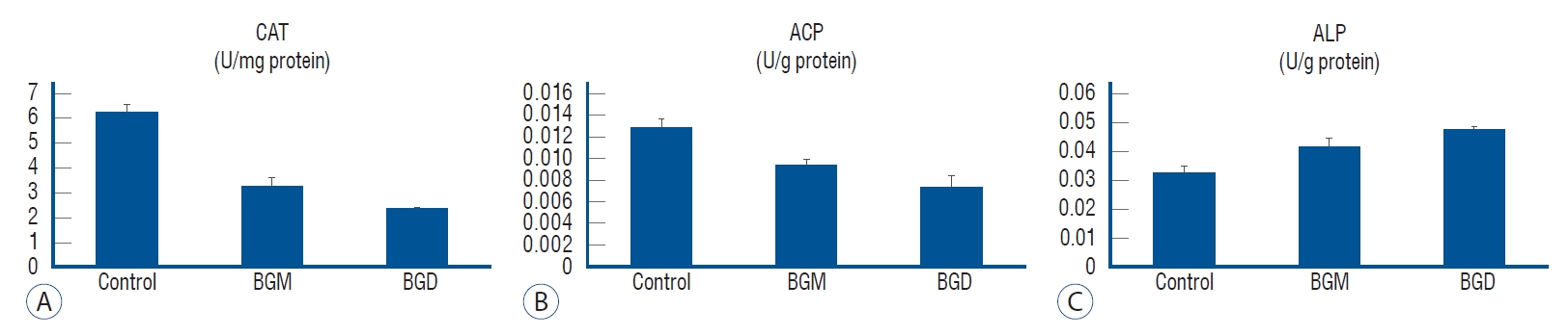
It was observed that the CAT enzyme activity significantly decreased in the groups where both BGM and BGD were administered 48 hours IC50 doses (6.6 and 26 mM, respectively) compared to the control (p<0.01) (Fig. 3A). Acid phosphatase (ACP) enzyme activity decreased in BGM and BGD groups compared to the control (p<0.05) (Fig. 3B). The increase in alkaline phosphatase (ALP) enzyme activity in BGM and BGD groups was not statistically significant when compared with the control (p>0.05) (Fig. 3C).
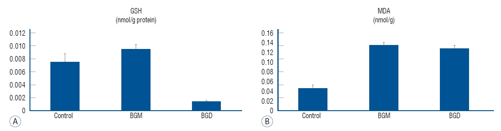
While the increase observed in BGM in total GSH amount was not statistically significant (p>0.05), a significant decrease was observed in the BGD group (p<0.001) (Fig. 4A). The increase in the amount of lipid peroxidation metabolite MDA in both boron-derived groups (p<0.05) indicates an increase in lipid peroxidation in both groups (Fig. 4B).
Antimicrobial effect of BGM and BGD on U87MG GBM cells
The antimicrobial activity results determined by the disk diffusion method are given in Table 1. According to these results; It was determined that the IC50 dose (6.6 mM) detected according to the results of the MTT test of the BGM had an antibacterial effect against S. aureus ATCC 29213 (11 mm) and E. coli ATCC 25922 (14 mm) strains but had no antifungal effect against C. parapsilopsis ATCC 22019 strain. Similarly, it was determined that the IC50 dose (26 mM) detected according to the results of the MTT test of the BGD had an antibacterial effect against S. aureus ATCC 29213 (8 mm) and E. coli ATCC 25922 (11 mm) strains but had no antifungal effect against C. parapsilopsis ATCC 22019 strain.
DISCUSSION
Boron-containing compounds are a potential agent for treatment against recurrent malignant cancers [27]. Boron compounds have been found to be effective against leukemia, breast cancer, lung cancer, prostate cancer, and ovarian cancer [10]. We chose BGM and BGD cytotoxic agents, we used in our study because of their anti-cancer effects.
Çelik et al. [11]. The cytotoxic effect of borax pentahydrate on the U-87 MG cell line was observed both at an increasing concentration (1, 10, 100, 500, 1000, 2500, 5000, 7500, and 10000 µM) and time (24, 48, and 72 hours) in the study they analyzed, they determined that borax pentahydrate had an effect on cell viability, especially after the 24th hour, depending on the dose and time and calculated the IC50 value at the 72nd hour as 2454 µM. Other researchers found the IC50 value of 17 mM as a result of applying different doses of boron (2.5, 25, and 50 mM) to GBM cells [5]. Meiyanto et al. [27] performed boron compaund in different cancer types (MCF-7/HER-2, MCF-7, RAW 264.7, and 4T1 with IC50 value of 12 µM, 54 µM, 26 µM, and <10 µM, respectively) According to their study, they found that the development of the new CCB-2 compound containing boron they synthesized as an anti-cancer agent is promising [27]. In our study, according to the statistical analysis after application of 500 µM, 1 mM, 5 mM, 10 mM, 25 mM, and 50 mM BGM and BGD to GBM cells for 48 hours, the IC50 value at 48 hours was calculated as 6.6 and 26 mM, respectively. Canturk et al. [10] found that administration of 1 mM boric acid for 48 hours in HL-60 cells reduced cell viability by 50%. In our study, we detected the survival rate in U87MG GBM cells treated with 500 µM, 1 mM, 5 mM, 10 mM, 25 mM, and 50 mM BGM was 106%, 102%, 61%, 51%, 33%, and 29%, respectively. Cell viability was determined as 90%, 88%, 85%, 82%, 77%, and 54%, respectively, by applying 500 µM, 1 mM, 5 mM, 10 mM, 25 mM, and 50 mM BGD to U87MG GBM cells. This result shows that boron compounds that we synthesized as the upper form of boron have a cytotoxic effect on U87MG GBM cells, especially BGM can be used in the treatment. These results show that BGM we synthesized is more effective than boron in GBM.
Cancer cells have high ROS production due to fast metabolism and impaired cellular signaling mechanism. High ROS levels are generally detrimental to cells, and the redox state of cancer cells is different from that of normal cells. Cancer cells, therefore maintain ROS levels above a low cytostatic level, but below levels that would be cytotoxic, at a moderately high tumorigenic level [17]. In the increase of ROS levels in cancer, the type of the produced radical, the region where the radical is located and the local concentration is important. ROS sensitive signaling pathways involved in cell growth/proliferation, differentiation, protein synthesis, glucose metabolism, cell survival, and inflammation in many types of cancer are also constantly increasing.
In cancer cells, higher than normal ROS production is balanced by an equal degree of antioxidant activity to maintain redox balance [13]. CAT, one of the powerful antioxidants in the cell, is the primary enzymatic defense system. H2O2 is metabolized by CAT into the water and molecular oxygen [17]. CAT, it is a primary enzyme of the antioxidant system in defense against oxidative stress that occurs in various cancer types. Rajneesh et al. [31] found that CAT levels increased in cancer patients. In our study, it was found that CAT enzyme level decreased as a result of the treatment of GBM cancer cells with boron derived compounds. These findings show that the complexes have properties to increase intracellular and intercellular oxidative damage. This situation has shown us that boron compounds can be effective against cancer by inhibiting the antioxidant defense system of cancer cells.
Acid and ALP, phospho-monoesterases belong to the enzyme family. ACPs are enzymes that transfer oxygen from water to inorganic phosphate in an acidic environment and catalyze the nonspecific hydrolysis of phosphate monoesters. ALP is an enzyme that catalyzes the hydrolysis of phosphate esters in an alkaline environment [12]. ALP is an enzyme responsible for the supply of phosphate groups required for cell cycle kinases. This feature is used as a biological marker in studies on cancer cells with the impaired cell cycle. However, in this study, ALP enzyme activity did not change in the GBM cells; while ACP enzyme activity reduced the effects of boron glycine compounds.
Kaynar et al. [21] examined antioxidant enzyme activities in cancer patients and found erythrocyte MDA, nitric oxide, total GSH levels, and erythrocyte SOD, CAT, and xanthine oxidase activities to be significantly higher than the control group. These studies have shown that there are significant changes in the antioxidant defense system caused by increased oxygen radicals in patients with cancer. The cancer cell detoxifies itself from ROS by increasing the level of antioxidant protein expression and ensures its safety. Thus, it is thought that a delicate balance of intracellular ROS levels is necessary for the function of the cancer cell. For example cancer cell can adapt to increased ROS level by increasing intracellular antioxidants such as GSH and heme oxygenase-1 [20]. In this study, while there was no significant change in Total GSH level in GBM cells treated with BGM, GSH level was decreased in the group treated with BGD. GSH depletion or down-regulation of GSH-related metabolic enzymes supports the restoration of active substance sensitivity in resistant cells. Therefore, the GSH antioxidant system plays an important role in the treatment of cancer. Given its potential as a target for the GSH antioxidant system, antitumor therapy, and reversal of drug resistance, it has recently become an attractive focus in cancer research [6,15].
Lipid peroxidation disrupts the function of the membrane and cell. The brain is more susceptible to oxidative stress than other tissues due to its low antioxidant enzyme activity and the high content of polyunsaturated fatty acids that can easily peroxid [38]. This situation supports the claim that unsaturated fatty acids in the membrane structure are most affected by lipid peroxidation. MDA measurement is most commonly done by the TBA method. It has been shown that the TBA method mainly measures the MDA itself in some experimental systems [42]. Since all measured MDA is a secondary product of lipid peroxidation, this method is thought to provide a reliable measure of lipid peroxidation in terms of MDA equivalents [32]. It has been observed that catechin and resveratrol increase lipid peroxidation in rat C6 glioma cells [35]. In this study, it was observed that the level of MDA in GBM cells treated with boron compounds was increased.
It is known that boron, boric acid, salts, and compounds have an important antimicrobial agent to affect yeast and bacterial infection. Researchers reported that boron has antimicrobial effects against C. albicans [29], S. aureus, P. aeruginosa, E. coli, Acinetobacter septicus, Aeromonas hydrophila, Brucella melitensis, Brucella abortus, Vibrio anguillarum, and Lactococcus garvieae [2,34,36,41]. Also, boron compounds come to the fore with their antimicrobial effects in new drug development studies due to the increasing antibiotic resistant strains. Since that situation is thought to affect other properties, the antimicrobial effects of those boron-containing compounds have been tested in addition to their anticancer and antioxidant effects.
The disc diffusion method was used to perform the evaluation of antimicrobial activities in vitro. It was determined in the study that BGM showed a higher antibacterial effect against test strains than BGD. BGM showed the highest antimicrobial activity among the test bacteria against E. coli ATCC 25922, a gram negative bacterium. IC50 dose of both compounds had no antifungal effect on C. parapsilopsis ATCC 22019. Yılmaz [41], in his study investigating the antimicrobial effects of boric acid and sodium tetraborate, which are boron compounds, found that these compounds have antibacterial effects on S. aureus and E. coli, similar to our study. On the other hand, while BGM and BGD we used were ineffective on P. aeruginosa, boric acid, and sodium tetra borate proved to be antibacterial.
Study limitations
In our study, the use of a commercial cell line instead of the primary cell line as the study subject, the absence of a 3D culture study with cells such as macrophage and endothelium to reflect the microenvironment, and the lack of advanced molecular analyzes for more descriptive results are the limitations of the study.