INTRODUCTION
Anterior cervical discectomy and fusion (ACDF) is a common surgical method for treating patients with degenerative cervical spinal diseases. This procedure could effectively remove compressive lesions located in front of the cervical medulla or cervical nerve roots, maintain segmental lordosis, and recover the intervertebral disc height. Initially, an autologous bone graft from the iliac crest was used as a substitute after removal of the intervertebral disc; however, several alternative devices such as polyetheretherketone (PEEK) cages, carbon-fiber cages, and three-dimensional printed titanium cages have been developed to reduce donor site morbidity [8,16]. Among them, PEEK cages have been widely used to replace autologous bone grafts [10].
ACDF with stand-alone PEEK cage (SA PEEK cage) had some complications, such as pseudoarthrosis, graft expulsion or collapse, cage migration, and cage subsidence. Restoring disc height is important for patients undergoing ACDF surgery, and cage subsidence is the major complication that reduces disc height [21]. There is no consensus on the definition of cage subsidence. However, a disc height reduction of ≥2 mm or ≥3 mm between immediately after surgery and the last follow-up was defined as cage subsidence [21].
To date, several studies have reported the risk factors affecting postoperative cage subsidence after ACDF with SA PEEK cages. The risk factor for cage subsidence is the distance of the cage from the anterior vertebral rim, preoperative cervical kyphosis, cage location, over-distraction, and excessive endplate preparation [1,20].
The traditional concept of cage subsidence is an inappropriate indicator for evaluating the preservation of disc height and prognosis of ACDF surgery. This is because, first, cage subsidence was theoretically expected to decrease the intervertebral neural foramen and worsen radicular pain. However, it is uncertain whether cage subsidence after ACDF affects the clinical prognosis [10,20]. Cage subsidence was only evaluated based on the amount of cage deposited into the vertebral body. Cage subsidence is difficult to verify if the postoperative disc height is maintained compared to that before surgery. Although postoperative cage subsidence occurs, if there is enough intraoperative distraction during surgery, intervertebral foraminal height can be maintained and symptoms related to spinal stenosis can be mitigated by sufficient distraction. Therefore, based on the changes in intervertebral disc height rather than cage subsidence, we planned the study to consider patients with complications such as reduced total disc height compared to that before surgery.
Second, few studies on the risk factors of subsidence have been conducted in patients with osteoporosis. Several studies have been conducted to confirm the relationship between osteoporosis and cage subsidence; however, no significant correlation has been identified.
Dual-energy absorptiometry (DEXA) is a common diagnostic tool for osteoporosis [4]. However, DEXA often fails to reflect the severe degenerative spine of the elderly [6]. Additionally, in cervical spine surgery, the bone condition of the cervical spine cannot be measured directly using DEXA. Quantitative computed tomography (CT) is currently the only method that can directly measure the BMD of the cervical spine [12]. However, the maintenance cost of the equipment is expensive and is not widely used, making it inappropriate as a standard test method. We considered a way to effectively assess cervical bone quality without introducing new equipment preoperatively. The region of interest (ROI) value of measuring the Hounsfield unit (HU) in conventional CT could reflect the quality of the cervical bone to some extent.
In this study, we first evaluated whether the ROI values of conventional cervical CT are correlated with the lumbar DEXA BMD; if the ROI value of cervical bone was meaningful, and after classifying the cage decrement with the new criteria aforementioned, we evaluated the relationship with postoperative disc height decrement after stand-alone ACDF surgery.
MATERIALS AND METHODS
Patient demographics
The study protocol was approved by the Institutional Review Board of Pusan National University Yangsan Hospital, which waived the requirement for informed consent due to the retrospective nature of this study (IRB No. 05-2021-115). We retrospectively reviewed the medical records of 40 patients who underwent single-level ACDF using a stand-alone PEEK cage between January 2012 and November 2018. All patients were treated for single-level pathology of the sub-axial cervical level by an experienced surgeon. The mean age was 52 years (range, 24-73). The inclusion criteria were as follows : 1) patients with neck pain and radiculopathy lasting for 3 months based on concordant preoperative magnetic resonance imaging (MRI); 2) cervical myelopathy due to acute rupture of the cervical disc; 3) all patients underwent a minimum 1-year follow-up period postoperatively with proper radiological examinations performed in outpatient clinics, and 4) only cases showing C7 and T1 vertebral body in the preoperative cervical sagittal plane examination.
Surgical procedure
All patients underwent a standard Smith-Robinson anterior approach to the cervical spine. After adequate exposure of the operative lesions, a Casper cervical retractor was placed, and a discectomy was performed in the standard manner. For complete decompression, we removed the bilateral subtotal uncinate process, and osteophytes of the posterior part of the vertebral body with high speed electric drill and Kerrison punch. We performed the bilateral partial uncinectomy to remove the remnant osteophyte regrowth, even in patients with unilateral symptoms. And we proceeded the uncinectomy laterally until the nerve root was fully decompressed. Posterior longitudinal ligament was selected removed for the patients in which ruptured disc directly compressed the dura. After decompression, the adjacent cartilaginous endplate was carefully removed to avoid damage to the bony endplate. And we found the proper cage size by inserting a different sized trial cages. Appropriate sized was selected according to immobility of trial cages following distractor removal. The implant was inserted under intraoperative fluoroscopy guidance. In all cases, a standalone anchored PEEK cage (Cornerstone-SR®; Medtronic Sofamor Danek, Memphis, TN, USA) was inserted without anterior plate anchoring. All patients were instructed to wear a soft collar for 2 months, postoperatively.
Radiologic assessment
All radiologic assessments were performed by two independent neurological patients who were not involved in the study and blinded to all clinical information. Routine preoperative radiological examinations consisted of plain radiographs (standing anteroposterior, lateral neutral, lateral flexion, lateral extension, and bilateral oblique views), CT, and MRI.
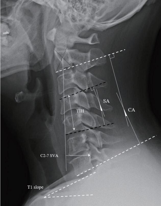
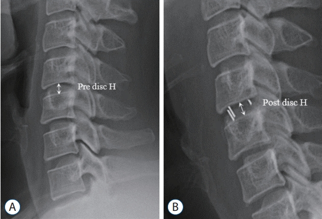
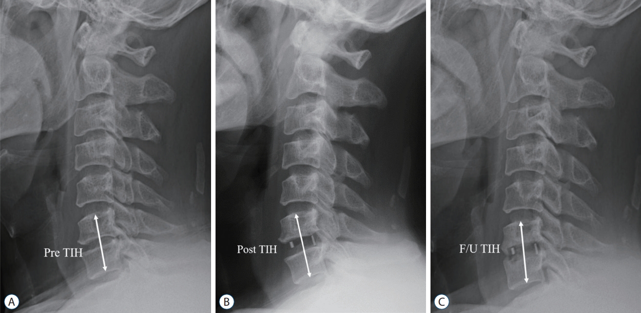
Radiologic parameters on plain radiographs included the C2-7 cobb angle (CA), T1 slope, C2-7 sagittal vertical axis (SVA), range of motion from C2-7, segmental angle (SA), total intervertebral height (TIH), and disc height. The C2-7 SVA, T1 slope, C2-7 CA, cage location, and intraoperative distraction were assessed on cervical neutral lateral radiographs in the free-standing position, with the patient’s head maintaining a horizontal gaze. Plain images were taken preoperatively and at postoperative day (POD) 1 and at 6 months, and 1 year postoperatively. TIH was defined as the distance from the midpoint of the upper endplate of the cephalic cervical vertebra to the lower endplate of the caudal cervical vertebra. Intraoperative distraction was assessed as the ratio of preoperative disc height to incremented disc height postoperatively. Cage decrement was defined as the reduction in TIH at the 6-month follow-up compared to preoperative TIH. Intraoperative distraction was measured as per the study by Barsa and Suchome [l1], and pseudoarthrosis was defined as segmental instability with a 2 mm alteration of the inter-spinous distance or a ≥2° increase in the SA [21]. All radiologic parameters were measured using commercial software (Marosis 5.0; INFINITT Healthcare, Seoul, Korea) and these measurement methods are presented in Figs. 1 and 2.
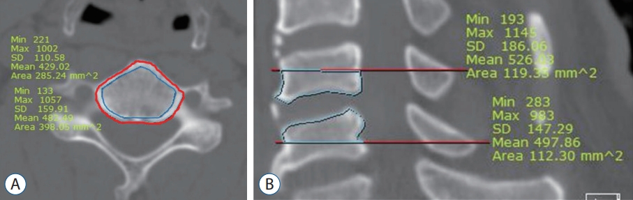
To assess the attenuation of the vertebral body, high-resolution CT scans of the cervical spine were retrospectively analyzed using a commercially available picture archiving and communication system (Somatom FORCE; Siemens, Forchheim, Germany). Two-dimensional reconstruction images were acquired in the axial and sagittal planes. CT attenuation was measured in HU by placing a click-and-drag elliptical ROI using a sagittal and axial CT image. HU values were calculated at the vertebra above and below the bone graft placement. In the sagittal image, HU values were measured in the lower half of the upper vertebra and the upper half of the lower vertebra at the midline. In the axial image, the HU value was measured in the mid-portion of the upper and lower vertebrae. To evaluate the effect of cortical bone thickness, HU values were measured by distinguishing the areas that contained the cortical bone. The measurement methods are shown in Fig. 3.
Statistical analysis
Statistical analysis was performed using SPSS (version 24.0; SPSS Inc., Chicago, IL, USA). Statistical significance was set at p<0.05. Normally distributed data of the groups were evaluated using the Student’s t-test and Mann-Whitney U test for parametric and nonparametric continuous variables, respectively. The Pearson correlation coefficient analysis was performed to confirm the relationship between continuous variables (radiologic parameters). A multiple logistic regression analysis was performed to determine the risk factors for subsidence. A receiver operating characteristic (ROC) curve analysis was used to determine the cut-off value, which was defined as the point corresponding to the maximum sum of the sensitivity and specificity.
RESULTS
The medical records and radiologic examinations of 40 patients were retrospectively analyzed. Our study included 19 male and 21 female patients aged between 24 and 73 years. The mean age of the patients was 52 years. Minimum followup period of all patients was 1 year and mean follow-up period was 18 months. Postoperative radiologic plain examination was taken at 1 day and at 6 months, and 1 year postoperatively.
For patients who underwent DEXA examination preoperatively, we analyzed the relationship between BMD values measured using DEXA and calculated HU values. The HU value showed a higher correlation with lumbar DEXA scores and a weak correlation with the T-score. However, it did not correlate with the femur BMD score. The HU value did not differ significantly depending on whether the cortical bone was included, and the HU value measured in the upper cortical bone was the most significant with DEXA BMD values. The results are summarized in Table 1.
Since the association of HU values and BMD has been verified, we used the HU values to assess the relevance of the subsidence.
Approximately 47.5% of the total patients were regarded as cage decrements. Cage decrement mostly occurred at the C5/6 level. However, there was no statistical relevance according to the surgical level, and no patient underwent revision surgery due to postoperative complications. We analyzed the clinical characteristics of patients with postoperative cage decrement. The demographic characteristics of the two groups are summarized in Table 2. No significant differences were found among clinical parameters. There were no differences in the preoperative radiologic parameters between the two groups.
In Table 3, we presented the radiologic change of the two groups. Regarding radiologic parameters, the cervical angle was almost unchanged, and there was no difference between the two groups. However, there were significant differences in the parameters related to intraoperative changes, such as variations in SA, TIH, and intraoperative distractions. There were also statistically significant differences in the parameters for measuring the HU value of the vertebral body at the surgery site. The HU value including cortical bone was approximately 70 units higher than that of the group that calculated only the cancellous bone. However, there was no difference in the results, even if the cortical bone was included. Among the areas for which the HU value was measured, the measurement of the upper vertebral body including cortical bone was the most significant area (p=0.011).
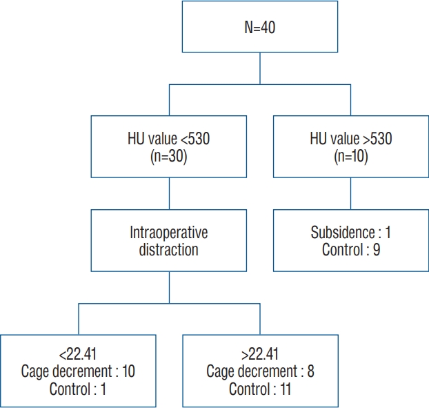
Using these identified factors, we performed an ROC curve analysis. Sensitivity and specificity were calculated for each cut-off value and the area under the curve (Table 4). The cutoff point was specified from the ROC curve using the optimal intersection of specificity and sensitivity. Based on the ROC curve, the cut-off point was 530 HU at the upper cortical and cancellous vertebrae (p=0.014; area under the curve [AUC], 0.727; sensitivity, 94.7%; specificity, 2.9%) and 22.41 HU at the intraoperative distraction (p=0.017; AUC, 0.722; sensitivity, 85.7%; specificity, 57.9%). Statistical significance was confirmed, except for lower cortical and sag lower HU values. Using this value, we converted these parameters into a bifurcated variable and assessed the multinomial regression analysis to evaluate the risk factors of postoperative decrement of ACDF. Finally, we confirmed that intraoperative distraction and HU value of the upper vertebral body were independent of postoperative subsidence. The flow chart for each of the two risk factors has been presented in Fig. 4.
If the patient had two risk factors, 90% of cage decrement after ACDF was predicted. The patient data was analyzed by applying the previous criteria defined by more than 3 mm cage subsidence after ACDF surgery and compared the composition of each group (Tables 5 and 6). Cage subsidence was present in 18 patients. In nine of 11 patients with two risk factors, cage subsidence occurred. A probability of 80% was able to predict the cage subsidence. The criteria for cage decrement did not differ significantly in patient composition compared to those with 3 mm cage subsidence.
A graph showing the variation in TIH according to the flow of time for each group involving risk factors has been presented in Fig. 5. A gentle slope was observed in patients with an HU value >530 compared to those with an HU value <530. Patients with adequate intraoperative distraction had no difference in the slope of the graph compared to the other patients. However, the final TIH was not reduced when compared to the preoperative TIH due to increased height.
DISCUSSION
ACDF using stand-alone PEEK is the most advantageous surgical strategy for cervical degenerative disease. With this procedure, we prevented the problem related to anterior cervical plating, including screw loosening, screw migration, and dysphagia due to soft tissue swelling [21]. However, according to the literature, there are many postoperative complications. Of various postoperative complications, graft subsidence belonged to major complications. Although there is some controversy over the criteria for postoperative subsidence, the incidence of graft subsidence after ACDF with stand-alone PEEK cage is reported to be 8.1-44.7% [2]. Although studies to identify the risk factors related to graft subsidence have been conducted, a consensus has not been achieved. As mentioned previously, there are several problems with the study of cage subsidence. Therefore, we introduce a new concept called cage decrement, focusing on whether the preoperative vertebral height is maintained. Concept of cage subsidence concentrated on the results that graft penetrated on the vertebral endplate, making it difficult to confirm whether preoperative height of neural foramen and vertebral height are maintained. In concept of cage decrement, the use of high-height cage acted as an independent variable contributing the increase of TIH. However, in concept of cage subsidence, the use of high-height cage acted as a dependent variable that exacerbates the cage subsidence. In Fig. 6, we introduced illustrated case. This case belonged to cage subsidence group, but it did not belong to cage decrement group. Although graft subsidence has been occurred, with use of the sufficiently high height cage, TIH and SA has been preserved.
As shown in Tables 5 and 6, classification by the new cage decrement criteria did not differ significantly from the previous classification based on the 3 mm cage subsidence criteria. However, when classified as per the 3 mm criterion, the multiple regression analysis did not produce statistically significant risk factors.
The identified risk factors in our study were the HU values and intraoperative distraction. The method of measuring the bone quality of the surgical lesion by checking the HU value in the operative lesion unit has been used in several studies. Schreiber et al. [15] introduced the HU measurement of each vertebral body using the standard PACS software. Unlike the lumbar spine, studies using the HU value of the cervical bone are lacking. Recent studies have evaluated the use of HU units of the commercial bone. Wang et al. [18] reported that lower preoperative CT HU values are associated with cage subsidence in single-level ACDF. Their study was similar to ours however, the following differences were noted. First, our research targeted patients who performed single-level ACDF without anterior plating. Moreover, the methods for measuring HU values were different. Second, we analyzed additional risk factors to forecast graft subsidence.
There are many studies on the association between the low HU values and mechanical failure in lumbar fusion surgery [13,14]. However, efforts to use HU values have been relatively limited in ACDF surgery and DEXA is not universally obtained by all spine surgeons before ACDF [5]. Our results showed that the cause of cage decrement depends on the bone quality and the HU value of conventional CT scans can reflect cervical bone quality. HU values also confirmed high consistency with lumbar BMD rather than that in femur heads and identified the usefulness of preoperative DEXA examination in cervical spine operation.
There is no consensus on how to measure the HU value of the vertebral body. Schneider et al. [15] first introduced the widely used measurement of HU values. The HU values of the cancellous bone of the vertebral body were measured at three axial image heights parallel to the endplates. However, the sagittal planes of the vertebral body were not measured. Lee et al.’s study [11] measured the vertebral body using sagittal and axial planes. Considering the characteristics of the cervical bone in which sclerotic changes of the vertebral body are frequently observed, we measured the vertebral body in three ways, including the cortical bone of the cervical bone. Although the most significant areas was the upper cortical vertebra including the cortical bone, statistical significance was also confirmed in other in other ways. We have a question why measuring the HU value in upper vertebral is the most valid site. According to the prior study about the trabecular bone density of cervical and lumbar vertebra [22], BMD value of high cervical vertebra is higher than BMD value of low cervical vertebra in normal human body. It can be assumed that cephalic cervical vertebra might reflect the quality of the entire bone better than caudal cervical vertebra, however additional study will be needed.
Intraoperative distraction has also been regarded as a cause of graft subsidence in some previous study [7,17,21]. In general, excessive intraoperative distraction caused a high static compressive force on the intervertebral endplate surface and potentially accelerated cage subsidence. However, it has a problem that PEEK cage with higher height should not be used. There was no objective parameters to use for determining the appropriate size during operation [7] and it is not easy to confirm whether the distractive force is appropriate or not. Adequate compressive force to endplate could be prevented the cage migration. And since our cases did not performed the anterior plate anchoring, we are free from the instrument failure such as screw back out or plate loosening caused by pull out force secondary to cage subsidence. In our study, we could confirm that cage decrement was most affected by the difference in HU values, that is, the cervical bone quality. Patients with relatively elevated disc height could eventually secure the disc space height and maintain lordosis of the SA. Intraoperative distraction is a factor that involves the surgeon’s discretion, and selecting a high-sized well-fitting graft could achieve a good radiologic outcome during ACDF surgery. However, excessive intraoperative distraction may reduce load transmission in the posterior column, worsening postoperative cervicalgia [3].
We conducted the retrospective study after defining a cage decrement based on POD 6 months. According to the prior study of cage subsidence after ACDF, the degree of cage subsidence was steepest during the first month after operation [1,8]. Afterward, the degree of cage subsidence gradually had slowed down until 6 months and the operative lesion has been stabilized after formation of fibrous union. Although there may be some exceptional cases, we set the criterion as POD 6 months in the study. Progression of cage subsidence was associated with degree of segmental instability (micro-motion) [9,19] and bony bridge formation [21]. Unfortunately, it is not feasible to perform CT examination on all cases. So, we measured the inter-spinous distance on flexion-extension lateral radiography and total pseudoarthrosis rates was 28%. This result was thought to be inferior compared to previously published result [18,21]. We thought that the main reason is absence anterior plate anchoring and excessive endplate damage during the end plate preparation.
This study has some limitations. First, it was a small retrospective study, and the follow-up period to confirm postoperative radiologic changes was short. And the age range of enrolled patients was diverse. So these aspects may reduce the confidence of the results. Second, our study introduced a new concept called cage decrement to accurately determine the degree of graft subsidence after ACDF. However, we have not evaluated comparative studies on whether these new concepts are clinically useful due to the limitation of a small number of patients. Third, we did not conduct a DEXA examination in all patients; it was performed in 27 out of 40 patients, which is a limitation in determining the usefulness of the DEXA examination.