| Home | E-Submission | Sitemap | Editorial Office |  
top_img
Journal of Korean Neurosurgical Society > Volume 65(5); 2022 > Article
Hwang, Chung, Kim, Yang, Choi, and Yoon: Value of Additional Instrumented Fusion in the Treatment of Thoracic Ossification of the Ligamentum Flavum

Abstract

Objective

The ossification of the ligamentum flavum (OLF) is one of the major causes of thoracic myelopathy. Surgical decompression with or without instrumented fusion is the mainstay of treatment. However, few studies have reported on the added effect of instrumented fusion. The objective of this study was to compare clinical and radiological outcomes between surgical decompression without instrumented fusion (D-group) and that with instrumented fusion (F-group).

Methods

A retrospective review was performed on 28 patients (D-group, n=17; F-group, n=11) with thoracic myelopathy due to OLF. The clinical parameters compared included scores of the Japanese Orthopedic Association (JOA), the Visual analogue scale of the back and leg (VAS-B and VAS-L), and the Korean version of the Oswestry disability index (K-ODI). Radiological parameters included the sagittal vertical axis (SVA), the pelvic tilt (PT), the sacral slope (SS), the thoracic kyphosis angle (TKA), the segmental kyphosis angle (SKA) at the operated level, and the lumbar lordosis angle (LLA; a negative value implying lordosis). These parameters were measured preoperatively, 1 year postoperatively, and 2 years postoperatively, and were compared with a linear mixed model.

Results

After surgery, all clinical parameters were significantly improved in both groups, while VAS-L was more improved in the F-group than in the D-group (-3.4±2.5 vs. -1.3±2.2, p=0.008). Radiological outcomes were significantly different in terms of changes in TKA, SKA, and LLA. Changes in TKA, SKA, and LLA were 2.3°±4.7°, -0.1°±1.4°, and -1.3°±5.6° in the F-group, which were significantly lower than 6.8°±6.1°, 3.0°±2.8°, and 2.2°±5.3° in the D-group, respectively (p=0.013, p<0.0001, and p=0.037). Symptomatic recurrence of OLF occurred in one patient of the D-group at postoperative 24 months.

Conclusion

Clinical improvement was achieved after decompression surgery for OLF regardless of whether instrumented fusion was added. However, adding instrumented fusion resulted in better outcomes in terms of lessening the progression of local and regional kyphosis and improving leg pain. Decompression with instrumented fusion may be a better surgical option for thoracic OLF.

INTRODUCTION

Ossification of the ligamentum flavum (OLF) is characterized by fibrosis, calcification and OLF, causing thoracic myelopathy [2,8,10]. Surgical decompression with or without fixation is recommended when thoracic myelopathy is symptomatic [20]. However, the need for instrumentation in thoracic OLF surgery is uncertain even though instrumented fusion surgery for thoracic ossification of the posterior longitudinal ligament (T-OPLL) is the standard treatment [24]. The symptoms of thoracic myelopathy usually improve after surgery, and the recurrence of OLF is rarely reported [3,14,30,44].
The causes of recurrence are not clear, but repeated mechanical stress on the residual ligamentum flavum after decompression-only surgery might be involved [12,18,36,40]. Because of this possibility, instrumented fusion has been advocated with decompression surgery [3,13,14,20,27,29,35,38,44]. Moreover, instrumented fusion may influence the local, regional and global sagittal spinal alignment, but studies analyzing post-surgical radiological changes are limited [1].
The purpose of this study was to compare the clinical and radiological outcomes of decompression only surgery with outcomes of decompression plus instrumented fusion for thoracic OLF.

MATERIALS AND METHODS

Patient populations

After approval from the Institutional Review Board at the Seoul National University Hospital (No. 1805-022-944) was obtained, we retrospectively reviewed the medical records of patients who had undergone surgery for thoracic myelopathy from January 2009 to December 2016.
Inclusion criteria were as follows : 1) patients who had undergone surgery for thoracic OLF, 2) patients who had followed up more than 24 months, 3) patients with preoperative, 12-month, and 24-month postoperative whole spine X-rays, and 4) patients with preoperative magnetic resonance (MR) imaging and computed tomography (CT) scan.
Exclusion criteria were as follows : 1) patients with a history of previous thoracic spinal surgery, 2) patients with combined herniated disc disease or ossification of the posterior longitudinal ligaments at the thoracic spine, 3) patients with myelopathic symptoms due to cervical lesions, and 4) patients who had undergone surgery with OLF in upper and mid-thoracic level (T2-8).
Finally, 28 patients were included in this study. Enrolled cases were divided into two groups; 17 patients with decompression only (D-group) and 11 patients with decompression plus instrumented fusion (F-group).

Surgical procedure and postoperative management

All operations were performed by experienced surgeons (CCK, JTA, and KCH) who had each performed more than 300 spinal surgeries.
Before 2013, decompression-only surgery was performed (D-group). The OLF and residual ligamentum flavum were removed after laminotomy, and the laminoplasty was performed with translaminar screws after decompression [31,41]. From 2013, instrumented fusions were routinely performed after decompression and removal of OLF (F-group). In the F-group, more than half of the facets were sufficiently removed to allow removal of the lateral parts of the OLF (Fig. 1), and instrumentations were performed only on the level of the OLF presence.
All patients were encouraged to ambulate from the day of surgery and were discharged 2-3 days after surgery. The patients were requested to wear a thoracic orthosis for a month in case of instrumented fusion.

Clinical evaluation

Mean age, sex proportion, body mass index (BMI), and the number of diabetic patients, symptom duration, follow-up duration, location of lesions were compared between two groups as preoperative characteristics. The mean follow-up period was 41.1±13.9 months (range, 25-74).
The Japanese Orthopaedic Association (JOA) scoring system was modified to evaluate the neurological function [42] (11, normal; 7-10, mild dysfunction; 4-6 moderate dysfunction; 0-3 severe dysfunction) (Table 1). All patients were requested to fill out questionnaires to evaluate the Korean version of Oswestry disability index (K-ODI) and Visual analogue scale of the back (VAS-B) and leg (VAS-L) [15]. To compare the clinical outcomes of the D-group and the F-group, we used the preoperative clinical parameters and those at postoperative 1 and 2 years.
In addition, complications from surgery and recurrence of OLF in the two groups during the follow-up period were investigated.

Radiographic evaluation

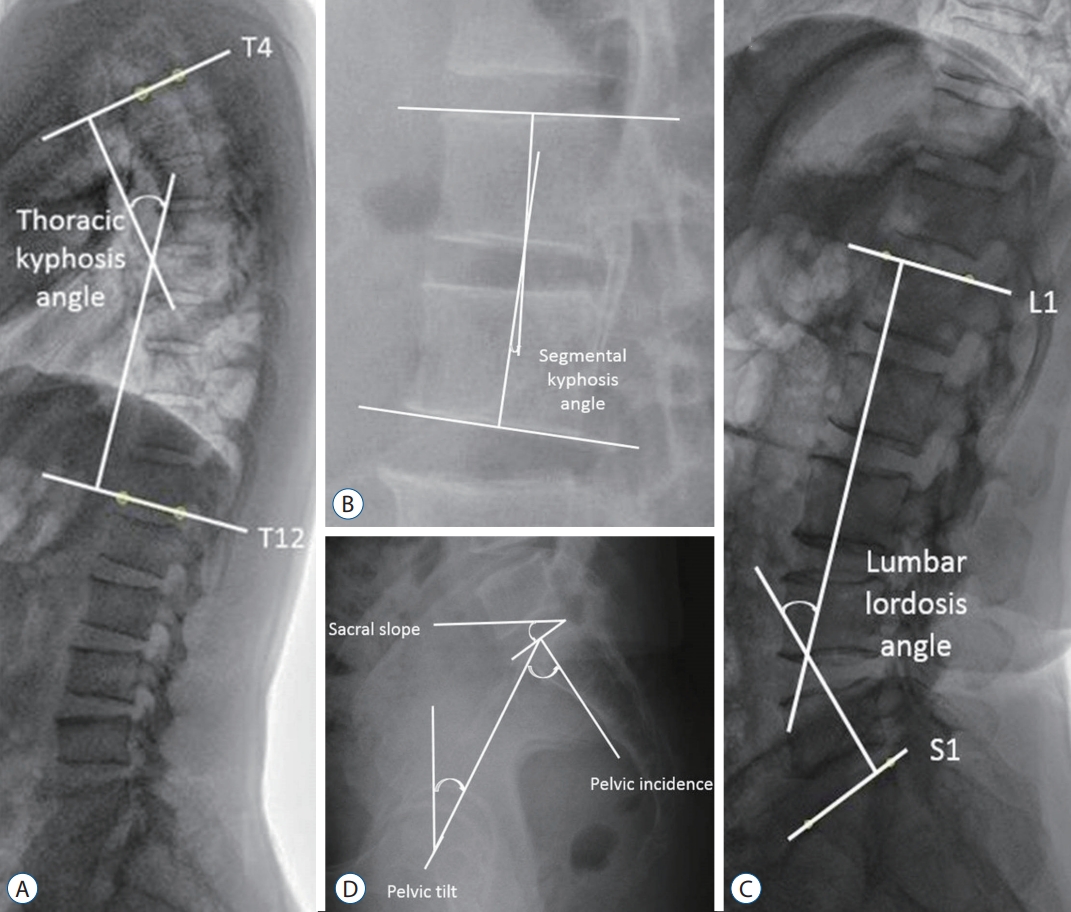
Whole-spine radiographs were taken in patients using 36-inch-long digital lateral radiographic films, with the arms of the patient flexed to 60° and the hips and knees fully extended preoperative, 12 months postoperative, and yearly thereafter. Patients were asked to stand up and look straight ahead during the whole-spine radiography. The following parameters were measured from the whole-spine radiographs; sagittal vertical axis (SVA), pelvic tilt (PT), sacral slope (SS), pelvic incidence (PI), thoracic kyphosis angle (TKA; T4-T12), segmental kyphosis angle (SKA) at the operative segment, and lumbar lordosis angle (LLA; L1-S1, a negative value implying lordosis) (Fig. 2). Cobb’s method was used to measure angles with positive values, meaning kyphosis.
Preoperative radiological parameters were compared with those at postoperative 1 and 2 years. All parameters were measured three times each at 2-week intervals by one neurosurgeon (SHH) and once by another neurosurgeon (JHY) blinded to the information of the patients using the tools in the picture archiving and communication system (Marosis, version 5483; Infinitt Healthcare, Seoul, Korea), which ran in a Microsoft Windows environment (Microsoft Corp., Redmond, WA, USA). For all radiological parameters, the intra-rater correlation coefficient was greater than 0.87 and the inter-rater correlation coefficient was greater than 0.81, and therefore the mean values of the four measurements were used for the present study.

Statistical analysis

Comparisons between continuous values were performed using Mann-Whitney U tests (or t-tests), while comparisons between non-continuous values were performed using Wilcoxon signed-rank tests and chi-square tests (or Fisher’s exact tests). A linear mixed model was used to compare parameters of the D-group to those of the F-group at admission, postoperative 1 year, and postoperative 2 years. Fixed effects included sex, age, operative methods, and parameters measured over periods, especially in order to control for the differences in the period between the D-group and the F-group. The correlations between the parameters were analyzed with Pearson correlation coefficient analysis. All statistical analyses were performed using SPSS (version 21.0; IBM Corporation, Armonk, NY, USA), and statistical significance was defined as p<0.05.

RESULTS

The preoperative characteristics of enrolled patients, clinical, and radiographic parameters are presented in Tables 2 and 3. Mean age, sex proportion, BMI, and the number of diabetic patients were not different between the groups, nor were any of their clinical parameters. Asymptomatic combined diseases (lesions without spinal cord compression) at other spinal levels were observed in 27 patients (multiple OLF, 17; diffuse idiopathic skeletal hyperostosis, 10; cervical OPLL, 6; ankylosing spondylitis, 1)
A gait disturbance with sensory change was the main presenting symptom in all patients (n=28). Other associated symptoms were back pain (n=23), leg pain (n=23), leg weakness (n=14), and voiding/defecation difficulty (n=12). After surgery, all clinical parameters were improved in both groups, and there was no difference in outcomes between groups, while the change in VAS-L was larger in F-group (Table 3). The radiological parameters were not different between the groups except for SVA (D-group, -1.1±3.9 mm vs. F-group, 3.5±5.4 mm; p=0.02).
Preoperative and postoperative 2-year SVA, pelvic parameters (PT, SS) and LLA were not significantly changed in either group. However, TKA and SKA were significantly changed after surgery in the D-group (p=0.02 and p=0.00, respectively), while they were not significantly changed in the F-group. The changes in TKA, SKA and LLA were significantly different between the groups. TKA, SKA, and LLA changed 6.8°±6.1°, 3.0°±2.8°, and 2.2°±5.3° after surgery in the D-group, while these changed 2.3°±4.7°, -0.1°±1.4°, and -1.3°±5.6° in the F-group (p=0.013, p<0.001, p=0.037, respectively) (Table 4). The change in SKA was correlated with that of TKA (r2=0.46, p=0.01), and the change in TKA was correlated with that of LLA (r2=0.33, p=0.01). Eventually, TKA was less kyphotic in the F-group than in the D-group (p=0.01), and LLA was more lordotic in the D-group than in the F-group after surgery (p=0.02) (Table 3).
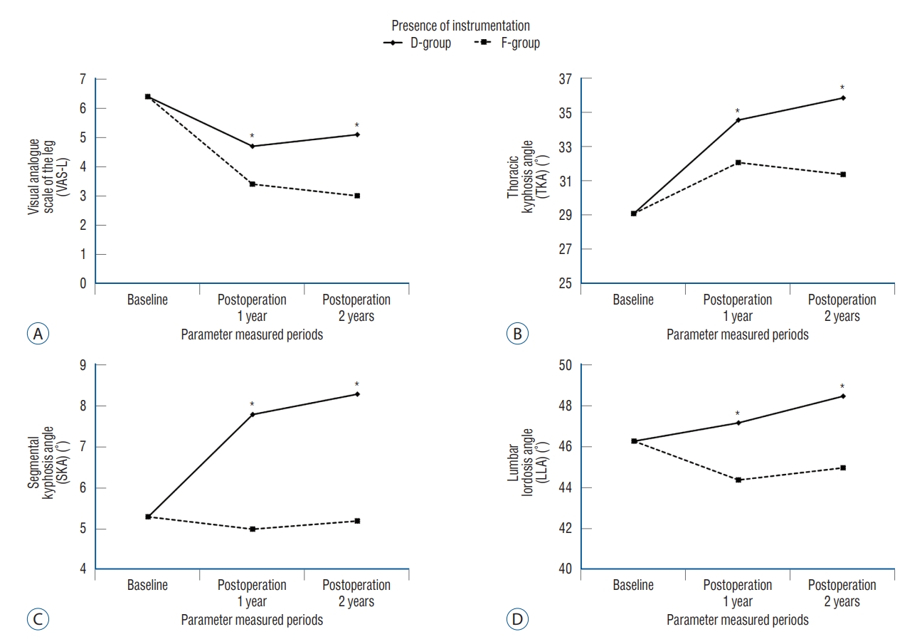
According to the linear mixed model analysis, VAS-L, TKA, SKA, and LLA showed significant differences between the two procedures (p=0.008, p=0.013, p<0.0001, p=0.037, respectively) (Table 4 and Fig. 3), but there were no differences in these parameters between following times.

Surgical complications and OLF recurrence

In the D-group, there was one patient who has undergone duroplasty due to intraoperative dural tear, one patient with asymptomatic exacerbation of OLF in the adjacent region, and symptomatic recurrence of OLF occurred in one patient at postoperative 24 months (Fig. 4).
In the F-group, there was one patient who has undergone duroplasty due to intraoperative dural tear, one patient who has undergone revision due to CSF leakage after surgery. There was no revision surgery or failure due to instrumentation during the investigation period.

DISCUSSION

OLF was first reported in 1920 by Polgar [32] and the prevalence of incidental OLF on chest CT is reported to be around 20% [16]. OLF is not only an isolated form of spinal column ossification but it also occurs in association with diffuse idiopathic skeletal hyperostosis, ankylosing spondylitis, and metabolic diseases such as Paget disease, hypoparathyroidism, and X-linked hypophophatemia [20]. However, since not all OLF patients are symptomatic, surgery is performed only in 0.6/10000 population [2]. Several studies have described the pathophysiology in the development and progression of OLF, including mechanical/traumatic stress [9,12,36], metabolic cause [23,34], diabetes mellitus [20], degenerative change [25], genetic/hereditary factor [7,17,37,43] and environmental factor [26,28]. In addition to these hypotheses, increased tensile stress on the ligamentum flavum has appeared as a more plausible pathophysiology [11,22]. Patients with OLF have a more kyphotic thoracic angle than the normal population [16]. Increased kyphosis may lead to stretching and increased tensile stress on the ligamentum flavum, and thus, it has been suggested as one of causative factors for OLF [16].
Because of the progressive nature of myelopathy in OLF, surgical decompression is recommended for symptomatic OLF, and the symptoms are mostly reversible with appropriate decompression [2,8,13,27]. However, the extent of decompression is still controversial. Because OLF usually progresses from the lateral to the medial part of the ligamentum flavum [35], removal of OLF from the medial to lateral part with preservation of the facet joint may leave the lateral part of OLF. This remained OLF may grow due to micro-motion and tensile stress at the operated segment [2,35], and the recurrence of OLF has been reported after decompression-only surgery [3,6,14,30,44]. To date, there has been no reported case of recurrence at the surgical site after instrumented fusion. To achieve sufficient decompression and total removal of OLF, at least half of the facet joint needs to be resected during decompression [9,45]. However, one study reported the progression of thoracic kyphosis after the removal of the facet joints and OLF [1]. Instrumented fusion enables sufficient decompression and eliminates mechanical factor/tensile stress contributing to the progression of OLF [9,12,19,21,36,45]. In addition to these effects on the operated site, we hypothesized that instrumented fusion might influence local, regional, and global spinal alignment.
In this study, we compared surgical outcomes between patients with decompression only (D-group) and patients with decompression plus instrumented fusion (F-group). Clinical outcomes were improved in both groups, but the improvement of leg pain was better in F-group. The cause of the better VAS-L in the F-group than in the D-group was not clear. The different extent of decompression between the two groups and the consequent improvement of VAS-L may be a plausible explanation. Observation of the two groups’ postoperative MR images and CT scans revealed that more than half of the facets were sufficiently removed to allow for removal of the lateral parts of OLF in the F-group. On the other hand, in the D-groups, the lateral parts of OLF were not sufficiently removed, as fewer than half of facets were removed.
According to results by Wang et al. [39] showed the JOA score improvement rate of the F-group at the 1-year follow-up was significantly different from that of the D-group. This might be because the spinal activity of the F-group had little interference with postoperative spinal cord recovery. These results showed that decompression with instrumented fusion had a significant clinical effect, especially for patients with OLF who had an extensive laminectomy [39].
In radiological outcomes, our study showed that global spinal alignment, which was represented by SVA and pelvic parameters, was not significantly changed and that there was no difference between the groups. Furthermore, the linear mixed model analysis, with which we expected to mitigate the difference in the period between the D-group and the F-group, revealed that the difference between the two groups was due to the surgical method as we expected but not the period of surgery.
Although our study showed that the surgery improved clinical parameters regardless of the use of instrumented fusion, the symptomatic progression of residual OLF occurred after the decompression-only surgery. In addition to its possible recurrence at the operated segment, we need to consider the development/progression of OLF at other levels. The progression of kyphosis at the operated segment and thoracic spine was evident in cases without instrumented fusion. Because OLF was observed on multiple levels in more than 50% of thoracic OLF patients, less TKA may be advantageous in reducing the development and progression of OLF [16].
Additionally, according to a large meta-analysis by Prablek et al. [33], the incidence of pseudoarthrosis in thoracic fusion is approximately 1.8%. Therefore, it is necessary to consider the deterioration of OLF due to excessive stress on the upper and lower adjacent joints.
Recently, a multicenter study reported that the posterior decompression and fusion with instrumentation method for thoracic OLF was only performed in 9.4% of patients from 2000 to 2008, but then the proportion increased to 48.9% from 2014 to 2017 in Japan [4]. Actually, the reason for this change may be that the techniques and instrumentation of spinal surgery have advanced markedly over the past 15 years [5]. However, the increase in the portion of instrumentation also reflects the current surgical trend toward posterior instrumented fusion surgery in thoracic OLF [4].

Limitation

The present study had several limitations. First, the use of instrumented fusion was not randomized and the design of the present study meant there was a chance of selection bias, different rehabilitation procedures might have influenced the results. Moreover, the small number of patients might have led to low statistical power. Second, although we showed improved SKA and TKA with instrumented fusion, their clinical significance may only be evident with a long-term follow-up study. Nonetheless, we showed that local and regional spinal alignments were improved by instrumented fusion. Third, the D-group and the F-group had different operation time points. To control for this, a linear mixed model was used, but the characteristics of patients before and after 2013, differences in postoperative care before and after 2013, and so on were impossible to control.
Even if the difference in time were controlled, the effect of the era would be included, so there could be a difference in effects between the two groups.

CONCLUSION

In thoracic OLF, the postoperative clinical outcomes were favorable regardless of whether instrumented fusion was added. However, the progression of kyphosis at both the index level and thoracic spine occurred after decompression surgery without instrumented fusion. Considering the pathophysiology of OLF, after enabling sufficient decompression, instrumented fusion may be appropriate. A long-term follow-up study with a large number of patients is necessary to show the effectiveness of the instrumented fusion.

Notes

Conflicts of interest

No potential conflict of interest relevant to this article was reported.

Informed consent

This type of study does not require informed consent.

Author contributions

Conceptualization : SHH, CKC; Data curation : SHH, JY; Formal analysis : SHH, YC; Funding acquisition : CKC; Methodology : CHK, YC; Project administration : SHH; Visualization : SHH; Writing - original draft : SHH; Writing - review & editing : CHK, CKC

Data sharing

None

Preprint

None

Acknowledgements

The study was supported by a grant of the Seoul National University Hospital (Nos. 3020210070 & 3020220200).

Fig. 1.
Computed tomography images showing the degree of facets removed to remove lateral parts of ossification of the ligamentum flavum (OLF) from the F-group and D-group. In the F-group (A and B), more than half of the facets were sufficiently removed to remove the lateral parts of OLF, but in the D-group (C), residual OLF was observed at the lateral part (arrows).
jkns-2021-0167f1.jpg
Fig. 2.
Measurements of radiographic parameters. Thoracic kyphosis angle (TKA) indicating the angle between the upper endplate of T4 and the lower endplate of T12 (A). Segmental kyphosis angle (SKA) indicating the angle between the upper and lower endplates at the disc level where surgery was performed (B). Lumbar lordosis angle (LLA) indicating the angle between the upper endplate of L1 and the upper endplate of S1 (C). Pelvic incidence (PI) indicating the angle between the line perpendicular to the sacral plate and the line connecting the midpoint of the sacral plate to the bicoxofemoral axis, sacral slope (SS) indicating the angle between the sacral plate and the horizontal plane, and pelvic tile (PT) indicating the angle between the lines connecting the midpoint of the sacral plate to the bicoxofemoral axis and the vertical plane (D).
jkns-2021-0167f2.jpg
Fig. 3.
Linear mixed-effects model graphs for VAS-L (A), TKA (B), SKA (C), and LLA (D) measurements over the periods with baseline correction for the D-group and F-group. The variable analyzed is the amount of change from the baseline. *p<0.05 for group analysis.
jkns-2021-0167f3.jpg
Fig. 4.
Recurrence case of ossification of the ligamentum flavum (OLF). A 43-year-old man underwent decompression surgery because of weakness in both legs (manual motor grade IV/V), decreased sensation, and voiding difficulty. Magnetic resonance (MR) imaging and computed tomography scan showed thoracic myelopathy due to OLF (A-C). The OLF was removed and laminoplasty was performed without instrumented fusion (D and E). Residual OLF was observed in the lateral part (D, arrows). Symptomatic thoracic myelopathy recurred at postoperative 2 years, and MR imaging revealed regrowth of the residual OLF (F).
jkns-2021-0167f4.jpg
Table 1.
Modified the Japanese Orthopedic Association scoring system for the assessment of thoracic myelopathy [42]
Neurological status Score
Lower-limb motor dysfunction
 Unable to walk 0
 Able to walk on flat floor with walking aid 1
 Able to walk up/downstairs with handrail 2
 Lack of stability & smooth reciprocation of gait 3
 No dysfunction 4
Lower-limb sensory deficit
 Severe sensory loss or pain 0
 Mild sensory deficit 1
 No deficit 2
Trunk sensory deficit
 Severe sensory loss or pain 0
 Mild sensory deficit 1
 No deficit 2
Sphincter dysfunction
 Unable to void 0
 Marked difficulty in micturition 1
 Minor difficulty in micturition 2
 No dysfunction 3
Table 2.
Characteristics of preoperative patients in D-group and F-group who had undergone surgery for thoracic ossification of the ligamentum flavum
Parameter Total (n=28) D-group (n=17) F-group (n=11) p-value
Age (years) 59.8±12.4 58.0±9.8 62.5±15.8 0.41
Female 14 (50.0) 10 (58.8) 4 (36.4) 0.26
BMI (kg/m2) 25.7±4.4 25.1±2.7 26.5±6.3 0.42
DM, yes 3 (10.7) 1 (5.9) 2 (18.2) 0.38
Symptom duration (months) 11.9±13.0 13.8±14.9 8.9±9.3 0.29
Follow-up duration (months) 41.1±13.9 44.5±15.1 34.8±9.0 0.11
Location of lesions
 T9-10 4 2 2
 T10-11 11 5 6
 T11-12 7 4 3
 T9-10-11 3 3
 T10-11-12 3 3
PI (°) 49.9±11.3 50.1±11.6 49.7±11.3 0.92

Values are presented as mean±standard deviation or number (%). Comparisons between continuous values were performed using Mann-Whitney U tests (or t-tests), while comparisons between non-continuous values were performed using Wilcoxon signed-rank tests and chi-square tests (or Fisher’s exact tests). D-group : decompression only surgery for thoracic ossification of the ligamentum flavum, F-group : decompression plus instrumented fusion surgery for thoracic ossification of the ligamentum flavum. BMI : body mass index, DM : diabetes mellitus, PI : pelvic incidence

Table 3.
Comparison of difference between preoperative and postoperative 2 years follow-up in each D-group and F-group, and comparison of differences preoperative and postoperative 2 years follow-up between D-group and F-group
JOA VAS-B VAS-L K-ODI SVA (mm) PT (°) SS (°) TKA (°) SKA (°) LLA (°)
D-group
Preoperative 5.9±1.3 5.2±2.1 5.8±2.3 20.6±9.7 -1.1±3.9 12.8±7.9 37.3±7.3 30.6±9.2 4.2±2.6 50.2±10.4
Postoperative 2 years 7.3±1.3 3.1±2.2 4.5±2.1 11.1±5.5 -0.4±2.9 12.3±7.5 37.7±6.5 37.4±6.5 7.3±2.9 52.4±9.4
p-value <0.01 0.01 0.1 <0.01 0.6 0.83 0.86 0.02 <0.01 0.52
F-group
Preoperative 5.2±1.3 6.5±1.4 6.9±1.6 24.4±7.1 3.5±5.4 13.7±7.9 36.0±7.0 27.5±9.4 6.3±4.0 42.3±10.2
Postoperative 2 years 6.8±1.0 4.2±1.8 3.5±2.4 14.5±6.1 3.3±6.0 14.8±8.3 35.0±8.2 29.8±6.5 6.6±3.3 41.0±14.1
p-value <0.01 <0.01 <0.01 <0.01 0.95 0.74 0.76 0.51 0.95 0.81
Between groups
Preoperative 0.18 0.08 0.19 0.28 0.02 0.79 0.65 0.39 0.06 0.057
Postoperative 2 years 0.26 0.18 0.28 0.14 0.03 0.40 0.34 0.01 0.55 0.02

Values are presented as mean±standard deviation or number. Comparisons between continuous values were performed using Mann-Whitney U tests (or t-tests). D-group : decompression only surgery for thoracic ossification of the ligamentum flavum, F-group : decompression plus instrumented fusion surgery for thoracic ossification of the ligamentum flavum. JOA : Japanese Orthopedic Association, VAS-B : Visual analogue scale of the back, VAS-L : Visual analogue scale of the leg, K-ODI : Korean version of the Oswestry disability index, SVA : sagittal vertical axis, PT : pelvic tilt, SS : sacral slope, TKA : thoracic kyphosis angle, SKA : segmental kyphosis angle at the operated level, LLA : lumbar lordosis angle (a negative value implying lordosis)

Table 4.
Comparison of changes from baseline in parameters between D-group and F-group using a linear mixed model
Postoperative 1 year Postoperative 2 years p-value* p-value
d_JOA 0.631 0.898
 D-group 1.5±1.0 1.5±0.9
 F-group 1.6±1.2 1.6±1.2
d_VAS-B 0.814 0.939
 D-group -2.2±1.5 -2.1±2.0
 F-group -2.2±2.1 -2.4±1.6
d_VAS-L 0.008 0.908
 D-group -1.7±1.7 -1.3±2.2
 F-group -3.0±2.6 -3.4±2.5
d_ODI 0.701 0.804
 D-group -9.7±8.6 -9.5±7.9
 F-group -10.9±7.6 -9.9±6.2
d_SVA (mm) 0.072 0.516
 D-group 1.7±3.1 0.6±2.3
 F-group -0.5±3.2 -0.2±3.5
d_PT (°) 0.806 0.152
 D-group 3.5±13.5 -0.6±2.8
 F-group 2.9±6.6 1.2±2.4
d_SS (°) 0.137 0.193
 D-group -0.8±4.4 0.4±2.9
 F-group -2.9±6.4 -1.0±2.5
d_TKA (°) 0.013 0.596
 D-group 5.5±6.1 6.8±6.1
 F-group 2.1±4.9 2.3±4.7
d_SKA (°) <0.001 0.602
 D-group 2.5±2.1 3.0±2.8
 F-group -0.3±1.4 -0.1±1.4
d_LLA (°) 0.037 0.509
 D-group 0.9±5.9 2.2±5.3
 F-group -1.9±4.8 -1.3±5.6

Values are presented as mean±standard deviation or number. D-group : decompression only surgery for thoracic ossification of the ligamentum flavum, F-group : decompression plus instrumented fusion surgery for thoracic ossification of the ligamentum flavum.

* Comparison between groups.

Comparison between postoperative 1 year and 2 years.

d_ : change from baseline, JOA : Japanese Orthopedic Association, VAS-B : Visual analogue scale of the back, VAS-L : Visual analogue scale of the leg, ODI : Oswestry disability index, SVA : sagittal vertical axis, PT : pelvic tilt, SS : sacral slope, TKA : thoracic kyphosis angle, SKA : segmental kyphosis angle at the operated level, LLA : lumbar lordosis angle (a negative value implying lordosis)

References

1. Aizawa T, Sato T, Ozawa H, Morozumi N, Matsumoto F, Sasaki H, et al : Sagittal alignment changes after thoracic laminectomy in adults. J Neurosurg Spine 8 : 510-516, 2008
crossref pmid
2. Aizawa T, Sato T, Sasaki H, Kusakabe T, Morozumi N, Kokubun S : Thoracic myelopathy caused by ossification of the ligamentum flavum: clinical features and surgical results in the Japanese population. J Neurosurg Spine 5 : 514-519, 2006
crossref pmid
3. Ando K, Imagama S, Ito Z, Kobayashi K, Ukai J, Muramoto A, et al : Progressive relapse of ligamentum flavum ossification following decompressive surgery. Asian Spine J 8 : 835-839, 2014
crossref pmid pmc
4. Ando K, Imagama S, Kaito T, Takenaka S, Sakai K, Egawa S, et al : Outcomes of surgery for thoracic myelopathy owing to thoracic ossification of the ligamentum flavum in a nationwide multicenter prospectively collected study in 223 patients: is instrumented fusion necessary? Spine (Phila Pa 1976) 45 : E170-E178, 2020
pmid
5. Bao H, Shu S, Yan P, Liu S, Liu Z, Zhu Z, et al : Fifteen years and 2530 patients: the evolution of instrumentation, surgical strategies, and outcomes in adolescent idiopathic scoliosis in a single institution. World neurosurg 120 : e24-e32, 2018
crossref pmid
6. Barath AS, Wu OC, Patel M, Kasliwal MK : Repeated recurrence of thoracic spine stenosis following decompression alone for ossification of the ligamentum flavum: case report. J Neurosurg Spine 30 : 332-336, 2018
crossref pmid
7. Cai HX, Yayama T, Uchida K, Nakajima H, Sugita D, Guerrero AR, et al : Cyclic tensile strain facilitates the ossification of ligamentum flavum through β-catenin signaling pathway: in vitro analysis. Spine (Phila Pa 1976) 37 : E639-E646, 2012
pmid
8. Chang UK, Choe WJ, Chung CK, Kim HJ : Surgical treatment for thoracic spinal stenosis. Spinal Cord 39 : 362-369, 2001
crossref pmid pdf
9. Chen J, Wang X, Wang C, Yuan W : Rotational stress: role in development of ossification of posterior longitudinal ligament and ligamentum flavum. Med hypotheses 76 : 73-76, 2011
crossref pmid
10. Guo JJ, Luk KD, Karppinen J, Yang H, Cheung KM : Prevalence, distribution, and morphology of ossification of the ligamentum flavum: a population study of one thousand seven hundred thirty-six magnetic resonance imaging scans. Spine (Phila Pa 1976) 35 : 51-56, 2010
pmid
11. Hur H, Lee JK, Lee JH, Kim JH, Kim SH : Thoracic myelopathy caused by ossification of the ligamentum flavum. J Korean Neurosurg Soc 46 : 189-194, 2009
crossref pmid pmc
12. Kaneyama S, Doita M, Nishida K, Shimomura T, Maeno K, Tamura Y, et al : Thoracic myelopathy due to ossification of the yellow ligament in young baseball pitchers. J Spinal Disord Tech 21 : 68-71, 2008
crossref pmid
13. Kang KC, Lee CS, Shin SK, Park SJ, Chung CH, Chung SS : Ossification of the ligamentum flavum of the thoracic spine in the korean population. J Neurosurg Spine 14 : 513-519, 2011
crossref pmid
14. Kanno H, Takahashi T, Aizawa T, Hashimoto K, Itoi E, Ozawa H : Recurrence of ossification of ligamentum flavum at the same intervertebral level in the thoracic spine: a report of two cases and review of the literature. Eur Spine J 27(Suppl 3):359-367, 2018
crossref pmid pdf
15. Kim DY, Lee SH, Lee HY, Lee HJ, Chang SB, Chung SK, et al : Validation of the korean version of the oswestry disability index. Spine (Phila Pa 1976) 30 : E123-E127, 2005
crossref pmid
16. Kim SI, Ha KY, Lee JW, Kim YH : Prevalence and related clinical factors of thoracic ossification of the ligamentum flavum-a computed tomographybased cross-sectional study. Spine J 18 : 551-557, 2018
crossref pmid
17. Kosaka T, Imakiire A, Mizuno F, Yamamoto K : Activation of nuclear factor kappaB at the onset of ossification of the spinal ligaments. J Orthop Sci 5 : 572-578, 2000
pmid
18. Li B, Guo S, Qiu G, Li W, Liu Y, Zhao Y : A potential mechanism of dural ossification in ossification of ligamentum flavum. Med hypotheses 92 : 1-2, 2016
crossref pmid
19. Li F, Chen Q, Xu K : Surgical treatment of 40 patients with thoracic ossification of the ligamentum flavum. J Neurosurg Spine 4 : 191-197, 2006
crossref pmid
20. Li Z, Ren D, Zhao Y, Hou S, Li L, Yu S, et al : Clinical characteristics and surgical outcome of thoracic myelopathy caused by ossification of the ligamentum flavum: a retrospective analysis of 85 cases. Spinal Cord 54 : 188-196, 2016
crossref pmid pdf
21. Liao CC, Chen TY, Jung SM, Chen LR : Surgical experience with symptomatic thoracic ossification of the ligamentum flavum. J Neurosurg Spine 2 : 34-39, 2005
crossref pmid
22. Maigne JY, Ayral X, Guérin-Surville H : Frequency and size of ossifications in the caudal attachments of the ligamentum flavum of the thoracic spine. Role of rotatory strains in their development. An anatomic study of 121 spines. Surg Radiol Anat 14 : 119-124, 1992
crossref pmid pdf
23. Matsui H, Katoh Y, Tsuji H : Untreated hypophosphatemic vitamin D-resistant rickets with symptomatic ossification of the ligamentum flavum. J Spinal Disord 4 : 110-113, 1991
pmid
24. Matsuyama Y, Sakai Y, Katayama Y, Imagama S, Ito Z, Wakao N, et al : Indirect posterior decompression with corrective fusion for ossification of the posterior longitudinal ligament of the thoracic spine: is it possible to predict the surgical results? Eur Spine J 18 : 943-948, 2009
crossref pmid pmc pdf
25. Miyazawa N, Akiyama I, Di Lorenzo N, Mouchaty H : Ossification of the ligamentum flavum of the cervical spine/comment. J Neurosurg Sci 51 : 139-144, 2007
pmid
26. Mobbs RJ, Dvorak M : Ossification of the ligamentum flavum: diet and genetics. J Clin Neurosci 14 : 703-705, 2007
crossref pmid
27. Moon BJ, Kuh SU, Kim S, Kim KS, Cho YE, Chin DK : Prevalence, distribution, and significance of incidental thoracic ossification of the ligamentum flavum in korean patients with back or leg pain : MR-based cross sectional study. J Korean Neurosurg Soc 58 : 112-118, 2015
crossref pmid pmc
28. Muthukumar N : Ossification of the ligamentum flavum as a result of fluorosis causing myelopathy: report of two cases. Neurosurgery 56 : E622, 2005
crossref pdf
29. Nishiura I, Isozumi T, Nishihara K, Handa H, Koyama T : Surgical approach to ossification of the thoracic yellow ligament. Surg Neurol 51 : 368-372, 1999
crossref pmid
30. Okada K, Oka S, Tohge K, Ono K, Yonenobu K, Hosoya T : Thoracic myelopathy caused by ossification of the ligamentum flavum. Clinicopathologic study and surgical treatment. Spine (Phila Pa 1976) 16 : 280-287, 1991
crossref pmid
31. Park SB, Jahng TA, Kim CH, Chung CK : Thoracic and lumbar laminoplasty using a translaminar screw: morphometric study and technique. J Neurosurg Spine 10 : 603-609, 2009
crossref pmid
32. Polgar F : Ueber intrartikulare wirbelvcerkalkung. Fortschr Geb Rontgenstr 40 : 292-298, 1920

33. Prablek M, McGinnis J, Winocour SJ, Reece EM, Kakarla UK, Raber M, et al : Failures in thoracic spinal fusions and their management. Semin Plast Surg 35 : 20-24, 2021
crossref pmid pmc
34. Sohn S, Yoon JW, Chung CK : Increased bone mineral density in patients with ossification of the ligamentum flavum: a case-control study. J Clin Densitom 17 : 195-199, 2014
crossref pmid
35. Trivedi P, Behari S, Paul L, Banerji D, Jain VK, Chhabra DK : Thoracic myelopathy secondary to ossified ligamentum flavum. Acta Neurochir (Wien) 143 : 775-782, 2001
crossref pmid pdf
36. Tsukamoto N, Maeda T, Miura H, Jingushi S, Hosokawa A, Harimaya K, et al : Repetitive tensile stress to rat caudal vertebrae inducing cartilage formation in the spinal ligaments: a possible role of mechanical stress in the development of ossification of the spinal ligaments. J Neurosurg Spine 5 : 234-242, 2006
crossref pmid
37. Uchida K, Yayama T, Cai HX, Nakajima H, Sugita D, Guerrero AR, et al : Ossification process involving the human thoracic ligamentum flavum: role of transcription factors. Arthritis Res Ther 13 : R144, 2011
crossref pmid pmc
38. Van Oostenbrugge RJ, Herpers MJ, De Kruijk JR : Spinal cord compression caused by unusual location and extension of ossified ligamenta flava in a caucasian male. a case report and literature review. Spine (Phila Pa 1976) 24 : 486-488, 1999
pmid
39. Wang ZW, Wang Z, Zhou YH, Sun JY, Ding WY, Yang DL : Clinical effect analysis of laminectomy alone and laminectomy with instrumentation in the treatment of TOLF. BMC Musculoskelet Disord 22 : 667, 2021
crossref pmid pmc pdf
40. Xu P, Wang C, He H, Xu G, Ye X : Spinal balance failure: a potential cause of spinal ligament ossification. Med hypotheses 76 : 908-910, 2011
crossref pmid
41. Yang SH, Kim CH, Chung CK, Park SB, Sohn S, Lee S : Bone fusion rate in the thoracic and lumbar spine after laminoplasty with laminar screws. Spine (Phila Pa 1976) 39 : E1325-E1330, 2014
crossref pmid
42. Yang Z, Xue Y, Zhang C, Dai Q, Zhou H : Surgical treatment of ossification of the ligamentum flavum associated with dural ossification in the thoracic spine. J Clin Neurosci 20 : 212-216, 2013
crossref pmid
43. Yayama T, Uchida K, Kobayashi S, Kokubo Y, Sato R, Nakajima H, et al : Thoracic ossification of the human ligamentum flavum: histopathological and immunohistochemical findings around the ossified lesion. J Neurosurg Spine 7 : 184-193, 2007
crossref pmid
44. Yonenobu K, Ebara S, Fujiwara K, Yamashita K, Ono K, Yamamoto T, et al : Thoracic myelopathy secondary to ossification of the spinal ligament. J Neurosurg 66 : 511-518, 1987
crossref pmid
45. Yu S, Wu D, Li F, Hou T : Surgical results and prognostic factors for thoracic myelopathy caused by ossification of ligamentum flavum: posterior surgery by laminectomy. Acta Neurochir (Wien) 155 : 1169-1177, 2013
crossref pmid pdf
Editorial Office
1F, 18, Heolleung-ro 569-gil, Gangnam-gu, Seoul, Republic of Korea
TEL: +82-2-525-7552   FAX: +82-2-525-7554   E-mail: office@jkns.or.kr
About |  Browse Articles |  Current Issue |  For Authors and Reviewers
Copyright © Korean Neurosurgical Society.                 Developed in M2PI
Close layer