INTRODUCTION
Vertebral compression fractures are the most common type of osteoporotic fractures, with approximately 1.4 million cases per year noted worldwide [6]. Vertebral compression fractures can result in severe pain, an inability to perform activities of daily living, and a marked reduction in quality of life [24]. Moreover, these fractures are associated with increased age-adjusted mortality [21]. To reduce this burden, evidence-based prevention and management are essential [1,20].
Over the past few years, percutaneous vertebroplasty (PVP), a minimally invasive technique, has been widely used worldwide to treat painful osteoporotic vertebral compression fractures (OVCFs) [9,19,25]. However, the effect of percutaneous cement augmentation are unclear, as are the benefits of PVP (including pain control, hospitalization period, quality of life, and vertebral body height restoration) and its adverse procedure-related events and incidence of adjacent compression fractures [7,8,12-15,18,22,26]. Conservative treatment may also benefit patients; hence, comparing the effects of conservative and PVP treatments on clinical and radiologic outcomes is essential. However, no concrete guidelines exist on how to administer conservative treatment in terms of medication, duration of bed rest, and use of orthoses.
This study compared the clinical and radiologic outcomes of PVP and conservative therapy using transdermal fentanyl patches (TFPs) in patients with OVCFs [2].
MATERIALS AND METHODS
Study design and patient groups
The Institutional Review Board of Gangneung Asan Hospital approved this study (approval number : 2019 05 005 GNAH IRB). This study included patients who were diagnosed with recent OVCFs and were treated conservatively using TFP or with PVP by two different spinal surgeons, according to the different treatment approaches, from March 2013 to December 2017.
Patients were included if they had single-level OVCFs, no history of treatment for vertebral fractures, no current medical comorbidity, and had been followed-up for at least 1 year with periodic evaluation of spine X-rays (anteroposterior/lateral views).
Patients were excluded if they had neurological deficits, pathological fractures, or unstable vertebral fractures involving the middle or posterior column of the spine.
Finally, 75 patients who underwent conservative treatment using TFPs and 56 patients who underwent PVP with a visual analog scale (VAS) score of 5 or more after 3 weeks of prevalent conservative treatment were included.
Statistical analysis included the student’s t-test, Mann-Whitney U test, and Fisher’s exact test, and a p-value of less than 0.05 was regarded as significant.
Treatment protocols for the conservative treatment and TFP groups
All patients underwent magnetic resonance imaging and assessment of bone mineral density (BMD) at admission and began osteoporosis medication and wore a corset for at least 3 months. After admission, a low-dose (12.5 µg) TFP was administered. Patients were prescribed combination tablets of tramadol (37.5 mg) and acetaminophen (325 mg) or acetaminophen (250 mg), ibuprofen (200 mg), and codeine phosphate (10 mg) as pro re nata oral medications.
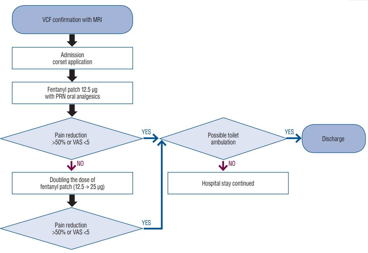
After the low-dose (12.5 µg) fentanyl patch application, the patients were asked whether their pain VAS score had changed. If the VAS score had decreased to below 5 or was 50% below the initial score, we continued to use the low-dose fentanyl patch for 1 month. However, if the patient complained of sustained pain with a VAS score that was more than 5 or 50% above the initial score, we increased the dose of the fentanyl patch by 25 µg. The fentanyl patches and corsets were applied simultaneously and immediately after admission. All patients were encouraged to use the toilet by walking with or without assistance, according to their pain tolerance. Absolute bed rest was not recommended, and discharge was recommended when toilet ambulation was possible and the VAS score was under 5 (Fig. 1).
Treatment protocol for the PVP group
In the PVP group, patients with sustained pain with a VAS score of more than 5 after 3 weeks of conservative treatment underwent PVP. Of the 170 patients with compression fractures, 56 (32.9%) were included and underwent PVP, based on the aforementioned guideline. This conservative treatment was performed with a non-steroidal anti-inflammatory drugs, COX-2 inhibitors, or combination tablets (tramadol and acetaminophen). Early ambulation was also encouraged after a brace was worn after identifying pain tolerance.
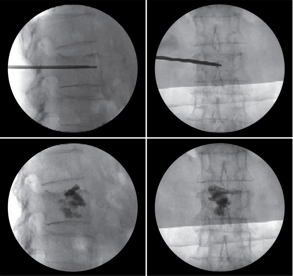
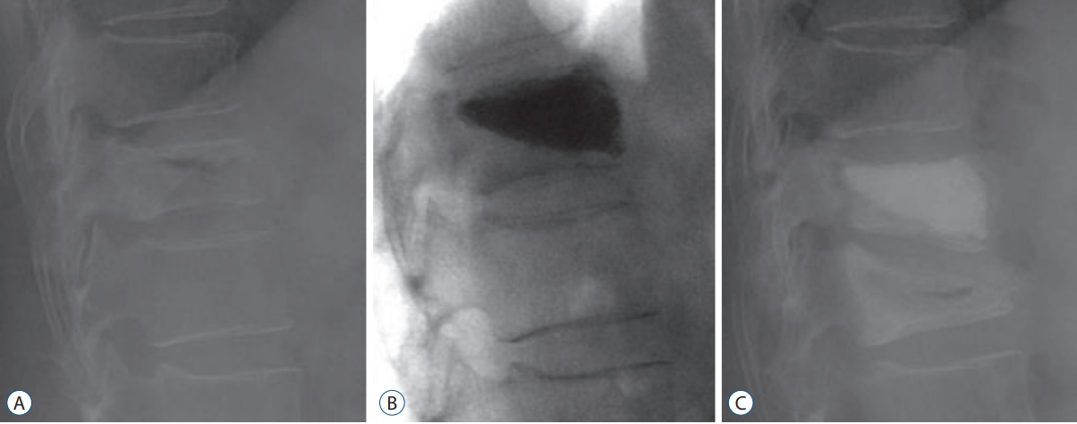
A vertebroplasty needle was inserted using a unilateral transpedicular approach. The liquid and powder components of polymethylmethacrylate were mixed, and the cement was injected through the pedicle under continuous fluoroscopic monitoring in the lateral view, with close attention paid to the posterior margin of the vertebral body and the epidural space. Postural reduction was also performed during PVP (Fig. 2).
Patients were discharged from the hospital after they attained tolerable toilet ambulation.
Comparison of baseline characteristics and patient outcomes
The two groups were compared for factors including age, sex, fracture level, and T-score of BMD (Table 1). The back pain VAS score was evaluated and compared at onset as well as 3 weeks and 1, 3, 6, and 12 months after the injury.
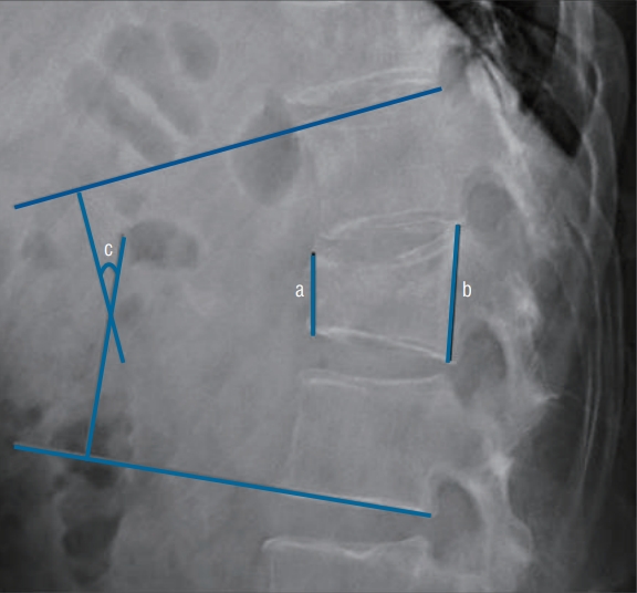
Radiographs, including anteroposterior and lateral X-ray images, were taken and compared at onset and 6 and 12 months after the injury. The compression rate was determined and compared by measuring the ratio of the anterior and posterior heights of the fractured vertebra level. The vertebral body wedge angle (kyphotic angle) was determined and compared by measuring the angle between the superior endplate of the vertebral body above and the inferior endplate of the vertebral body below the fractured vertebra on lateral X-rays (Fig. 3). The development of compression fractures adjacent to the index vertebra during follow-up was checked and compared.
RESULTS
The patients in TFP group were carefully observed for side effects of the fentanyl patch, such as nausea, vomiting, or dizziness. Such side effects developed in 17 patients (22.6%), even after the use of a low-dose (12.5 µg) fentanyl patch. We subsequently removed the patch from their medication and instead administered only oral analgesics.
In 16 patients (21.3%) whose pain was not controlled with the low-dose (12.5 µg) fentanyl patch, a higher dose of fentanyl (25 µg) was used. Six of the 75 patients underwent PVP because of insufficient pain reduction and low overall satisfaction.
Comparison of clinical outcomes between the TFP and PVP groups
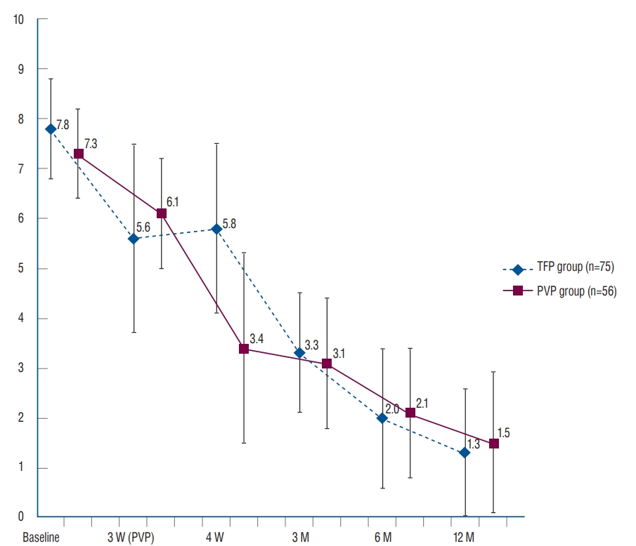
During the 12-month follow-up period, overall VAS scores improved over time in both groups (from 7.8 to 1.3 in the TFP group, and from 7.3 to 1.5 in the PVP group). However, no statistical difference was observed between the two groups when changes were compared from the baseline to 12 months (6.5 in the TFP group, 5.8 in the PVP group, p=0.62). The mean VAS scores of the TFP and PVP groups were 5.6 and 6.1, respectively, at 3 weeks (p=0.355). However, a significant difference in the VAS score was observed between the two groups at 4 weeks (1 week after PVP) (VAS score, 5.8 in the TFP group; VAS score, 3.4 in the PVP group; p=0.022) (Fig. 4).
Comparison of radiologic outcomes between the TFP and PVP groups
The compression rates in the TFP and PVP groups were 24.8 and 32.9, respectively, at admission. The baseline compression rate was significantly higher in the PVP group (p=0.001). After 12 months, the compression rates of the TFP and PVP groups were 31.9 and 28.1, respectively. Although kyphosis progressed in the TFP group, compression did not progress after kyphosis reduction in the PVP group (p=0.001).
The mean kyphotic angles in the TFP and PVP groups were 13.2° and 14.8°, respectively, at baseline (p=0.002). The mean kyphotic angles after 12 months differed significantly, at 12.4° and 13.6° in the TFP and PVP groups, respectively (p=0.013).
Five patients in the PVP group experienced adjacent compression fractures within 12 months, which differed significantly from the TFP group, in which none were observed (p=0.013) (Table 2). Adjacent compression fractures occurred within 1 month in two patients (L1 L2, T11 T12), within 3 months in two patients (T10 T9, T12 L1), and within 1 year in one patient (L1 L2) (Fig. 5).
DISCUSSION
Although PVP has become the preferred method of treatment, its relative benefits for OVCFs remain unclear [7,10,16]. For example, a randomized controlled trial showed that pain relief and quality of life did not differ significantly between patients who underwent PVP and those who received conservative treatment [7]. Moreover, a recent study found that most patients treated conservatively had favorable clinical results26), and our study yielded similar results; although pain relief was greater in the PVP group 1 month after onset of OVCFs (1 week after PVP), the two groups subsequently showed similar pain relief.
Various imaging modalities have yielded varying results in assessing factors such as the compression ratio and kyphotic angle [26]. In the present study, the compression ratio increased after 12 months in the TFP group but decreased after 12 months in the PVP group. Because PVP involves postural reduction, kyphosis reduction is maintained for at least 12 months (31.9% vs. 28.1%, p=0.001). The initially significantly worse compression rate in the PVP group might have been due to the study including only patients for whom the initial 3 weeks of conservative treatment failed (24.8% vs. 32.9%, p=0.001).
However, the kyphotic angle improved in both groups over time. Although the between-group difference in the change in the kyphotic angle was statistically significant, the actual difference was less than 1°, suggesting that this difference may not be clinically important and may have been due to measurement bias.
Several adverse effects have been associated with vertebral augmentation, including cement leakage and adjacent segment fractures [5]. The complications of cement leakage include neurologic deficits and pulmonary embolisms. Moreover, the incidence of new OVCFs after vertebroplasty has been reported to range from 8% to 52%23), with 41-69% of new vertebral compression fractures being immediately adjacent to the treated vertebra [8,11,14,15,18,22].
Five of our patients (8.9%) who underwent PVP, but none in the TFP group, were diagnosed with adjacent compression fractures within 1 year and mostly within 3 months. Previous studies have also shown that most new OVCFs occur within 3 months after PVP [8,11,14,15,18,22].
Although age differed significantly in the present study (8.1 years, TFP group, 63.4 years old vs. PVP group, 71.5 years old), BMD was similar in the two groups (p=0.751). Because the PVP group included only patients for whom the initial 3 weeks of conservative treatment failed, younger patients might have been excluded because of their better response to traditional conservative treatment. Collectively, the results indicate that conservative treatment might be more effective for OVCFs. In addition, we cannot exclude the possibility that more adjacent fractures occurred because of the older age in the PVP group rather than because of the direct stress effect of cement augmentation.
The reason for BMD scores being similar despite the significant difference in age between the two groups is believed to be the rapid decrease of BMD that generally occurs before the early 60s; a slow and gradual decrease occurs from the mid60s to early 70s, resulting in little difference in BMD scores in our results [3].
Generally, fentanyl patches have some of the same adverse effects as other opioids, mainly sedation, hypoventilation, nausea, vomiting, constipation, and urinary retention. In comparison with oral morphine, they cause fewer gastrointestinal adverse events. Because of the possibility of these side effects, we had to recommend that all patients be admitted to hospital and use individual fentanyl patches under careful monitoring. We could not administer fentanyl patches to 17 patients because of nausea and dizziness; however, no patients had a decrease in respiratory drive. Previously, we have shown the efficacy and safety of fentanyl patches for chronic pain [17]. Fentanyl patches offer an alternative to oral morphine, and their effectiveness and tolerability have been demonstrated in several trials [4]. In addition to safety and experience, the most crucial reason to choose fentanyl patches instead of other oral opioids is the convenience of changing them every 3 days. Considering that 32.9% of the patients (56 out of 170 patients) underwent PVP rather than popular and traditional conservative treatment, but only 8.0% (six out of 75 patients) underwent PVP in the TFP group, our initial TFP application seemed to be more beneficial in reducing the conversion rate from conservative treatment to PVP if we carefully monitored the development of side effects.
This study had several limitations. First, this study had a retrospective design and was not a randomized control trial. Second, it measured only pain and radiologic factors including compression rate and kyphotic angle, but did not assess other functional outcomes and other sagittal radiological parameters. Finally, this study was non-homogeneous, in that the two groups of patients were treated by two independent surgeons.
CONCLUSION
We compared clinical and radiological outcomes between the TFP and PVP groups. The immediate pain reduction effect was superior in the PVP group, but the final clinical outcomes were similar. Although the PVP group exhibited more preserved segmental alignment than the TFP group did for 1 year, the development of adjacent fractures was significantly higher. Although conservative treatment using TFPs seems to be beneficial in reducing the conversion rate from conservative treatment to PVP, the rate of the occurrence of side effects (in this study, 22.6%, 17 out of 75 patients) should be carefully monitored.