A Comprehensive Analysis of Potential Complications after Oblique Lumbar Interbody Fusion : A Review of Postoperative Magnetic Resonance Scans in Over 400 Cases
Article information

Abstract
Objective
This study focuses on identifying potential complications following oblique lumbar interbody fusion (OLIF) through routine magnetic resonance (MR) scans.
Methods
From 650 patients who underwent OLIF from April 2018 to April 2022, this study included those with MR scans taken 1-week post-operatively, and only for indirect decompression patients. The analysis evaluated postoperative MR images for hematoma, cage insertion angles, and indirect decompression efficiency. Patient demographics, post-operatively symptoms, and complications were also evaluated.
Results
Out of 401 patients enrolled, most underwent 1- or 2-level OLIF. Common findings included approach site hematoma (65.3%) and contralateral psoas hematoma (19%). The caudal level OLIF was related with less orthogonality and deep insertion of cage. Incomplete indirect decompression occurred in 4.66% of cases but did not require additional surgery. Rare but symptomatic complications included remnant disc rupture (four cases, 1%) and synovial cyst rupture (four cases, 1%).
Conclusion
This study has identified potential complications associated with OLIF, including approach site hematoma, contralateral psoas hematoma, cage malposition risk at caudal levels, and radiologically insufficient indirect decompression. Additionally, it highlights rare, yet symptomatic complications such as remnant disc rupture and synovial cyst rupture. These findings contribute insights into the relatively under-explored area of OLIF complications.
INTRODUCTION
Oblique lumbar interbody fusion (OLIF) provides indirect decompression and mechanical stability [6,12] while preserving posterior structures compared with posterior/transforaminal lumbar interbody fusion [3,10], offering a less lumbar plexus traumatic alternative to direct lateral interbody fusion [6,18,20]. As a recent surgical option [17,25,28], OLIF has garnered attention for its potential complications, which primarily include intraoperative injuries like psoas paresis, sympathetic plexus and vessel damage, and postoperative issues such as persistent pain and contra-lateral root symptoms [1,7,18,22,28]. Given that OLIF uses a retroperitoneal approach for indirect decompression, unrecognized complications may exist. Therefore, routine postoperative magnetic resonance (MR) scans are essential for all patients to better understand and address these potential risks, rather than confining MR scans only to symptomatic cases after surgery.
Postoperative MR scans provide information on complications such as cage position, insertion angle, retroperitoneal hematoma, psoas muscle hematoma, cage malposition related to contra-lateral root injury, and contra-lateral disc rupture. Information on insufficient decompression of grade 3 or higher can also be obtained [24]. In the present study, we only targeted patients who underwent indirect decompression. Moreover, we anticipate that multiple MR scans will improve the understanding of potential complications and the ability to detect them.
For over 3 years, our hospital has routinely conducted MR scans 1-week post-OLIF. With a repository of over 600 cases, we believe we have collected enough information to validate the potential postoperative findings. In this study, we aim to present the potential complications after OLIF identified on postoperative MR scans.
MATERIALS AND METHODS
The study protocol was approved by the Institutional Review Board of Pusan National University Yangsan Hospital which waived the requirement for informed consent due to the retrospective nature of this study (IRB No. 55-2023-031).
Inclusion and exclusion criteria
Between April 2018 and April 2022, we performed OLIF on 650 patients. The inclusion criteria were as follows : 1) patients with degenerative spondylosis accompanied by segmental instability that did not respond to drug therapy for more than 6 weeks; 2) patients who underwent MR scans 1 week after surgery, regardless of symptoms (from December 2019 onward); 3) patients who had OLIF performed from the L2 level to the S1 level; and 4) patients who underwent posterior percutaneous pedicle screw insertion.
The exclusion criteria were : 1) patients who could not undergo postoperative lumbar MR imaging due to postoperative internal medical conditions; 2) patients who had direct decompression (additional laminectomy performed using the posterior approach); and 3) patients who underwent OLIF at the L5/S1 level using the between bifurcation approach, rather than pre-psoas approach.
Operative technique
The OLIF procedure was conducted according to the previously reported method [18,26]. Herein, we describe certain surgical factors that are relevant to the outcome measures; cage insertion : throughout surgery, we maintained orthogonal maneuvers to the utmost extent possible. The cage was inserted until the middle radiopaque marker of the cage aligned with the spinous process of the respective level. We took care to ensure that the right cage marker did not exceed the midpoint of the right pedicle during cage insertion (Fig. 1A). Additionally, in the lateral image, we took precautions to ensure that the right posterior cage marker did not exceed the posterior margins of the vertebral bodies above and below (Fig. 1B). Drain insertion : regarding retroperitoneal hematoma, we did not place drains in any of the patients.
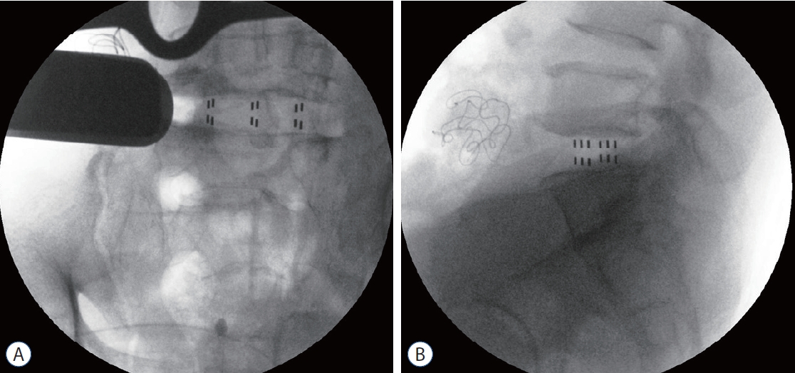
Landmarks for oblique lumbar interbody fusion cage insertion. A : Anteroposterior image : the middle radiopaque marker of the cage does not push beyond the spinous process. B : Lateral image : the most dorsal radiopaque marker of the cage does not surpass the posterior margin of the upper vertebral body.
Assessment of radiological and clinical outcomes
Postoperative MR images were obtained using either 1.5-Tesla or 3.0-Tesla MR imaging, and only T2 axial and T2 sagittal images were acquired. The following parameters were evaluated in the postoperative MR scans : 1) approach site hematoma and contralateral psoas hematoma : we checked for the presence of T2 high fluid collection (hematoma) up to the left retroperitoneal space (from the abdominal wall to the left vertebral body and left psoas muscle). Next, we checked for hematoma in the contralateral psoas muscle at the fusion level. The volume of the hematoma was categorized as none, scanty (<10 mL), or >30 mL (Fig. 2). We used the intra-cerebral hemorrhage volume measurement method to estimate hematoma volume. 2) Cage insertion angle and depth : using the T2 axial image, we measured the cage depth and angle at the disc level. Cage depth was determined by drawing an extension line following the cage’s posterior margin and measuring the distance to the right border of the body. The cage angle was measured as the Cobb angle between an imaginary line connecting both facet joints and the imaginary line passing through the center of the cage (red dotted lines) (Fig. 3). Clinically, deep cage insertion (defined as a depth exceeding 5 mm) was associated with remnant disc rupture to the contralateral neural foramen. 3) Indirect decompression : the evaluation, particularly of central canal stenosis, was conducted using the method by Schizas et al. [23]. Especially in cases of preoperative foraminal stenosis or spondylolisthesis with segmental instability, many patients did not have severe central stenosis, and in these instances, changes in central stenosis were not within the scope of our analysis. Thus, the effects of indirect decompression were only evaluated in patients whose preoperative stenosis severity was graded as “C” or “D.” We defined postoperative stenosis grades of “C” and “D” as insufficient indirect decompression. And 4) other findings : when comparing the preoperative and postoperative MR scans, we evaluated both the presence of remnant disc rupture and any unexpected findings.
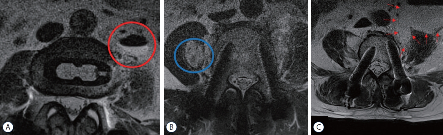
Evaluation of approach site or contralateral site hematoma. A : Red circle : approach site hematoma (more than 10 mL). B : Blue circle : contralateral site hematoma (scanty). C : Red arrows : approach site hematoma more than 30 mL.
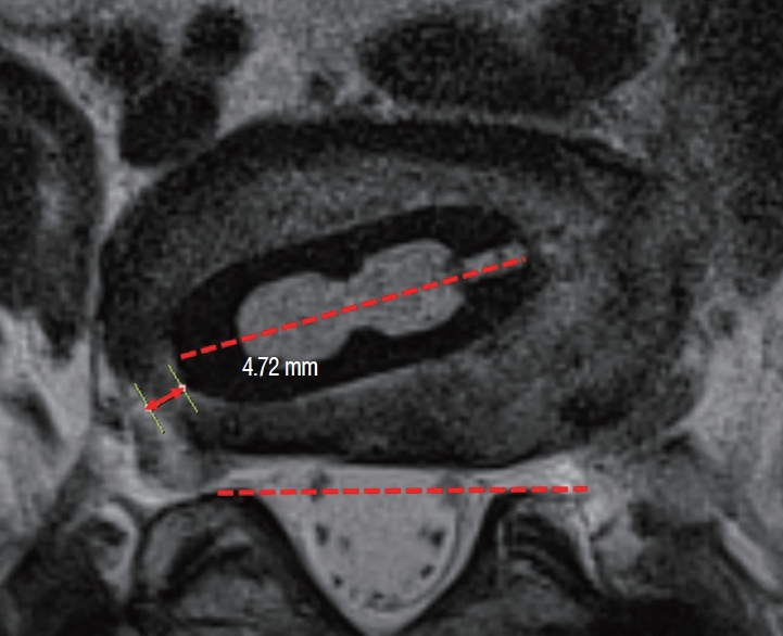
Measurement of cage insertion angle and depth (red arrow). Cage depth was determined by drawing an extension line following the cage’s posterior margin and measuring the distance to the right border of the body (red arrow : 4.72 mm). The cage angle was measured as the Cobb angle between an imaginary line connecting both facet joints and the imaginary line passing through the center of the cage (red dotted lines).
Clinically, we recorded patient age, gender, body mass index, bone mineral density, and American Society of Anesthesiologists physical status classification. Specifically, we collected data on symptoms that did not improve postoperatively, worsened symptoms, and newly developed pain or weakness. We evaluated postoperative symptoms in patients who experienced potential complications, and for some representative cases, we provided a case presentation. The muscle strength was measured using the Medical Research Council (MRC) scale for muscle strength.
RESULTS
In total, 401 patients who underwent postoperative MR imaging were enrolled. The characteristics of the patients and surgical details of the enrolled group are summarized in Table 1. As only patients who underwent indirect decompression were included, most were 1- or 2-level patients (50.9% and 36.7%, respectively), while 3- and 4-level patients constituted a smaller proportion (12.0% and 0.5%). For the same reason, most procedures were performed at the L3/4/5 levels.

Operative data
Approach site hematoma occurred in 65.3% of patients, with significant hematomas of more than 10 mL observed in 5.5% of the total. Notably, as the number of surgical levels increased, the frequency of hematomas larger than 10 mL also increased. No symptoms related to these hematomas were observed, and there were no subsequent infections or additional surgery. Contralateral psoas hematoma occurred in 19% of patients, and none exceeded 10 mL. No symptoms related to contralateral psoas hematoma were observed, and no correlation between multi-level surgery and hematoma was found (Table 2).

Approach site or contra- lateral site hematoma
Lower orthogonality was observed in the OLIF cage insertion at the caudal level. Notably, significantly lower orthogonality was observed at L4/5 than at L2/3 and L3/4, and this trend was even more pronounced at L5/S1. Cage insertion depth did not vary by level (Fig. 4). However, deep cage insertion occurred more frequently at lower levels (Table 3). In one case, the distance from the cage to the right neural foramen was 0 mm. fortunately, the patient only had mild right leg radiating pain, which allowed conservative treatment.
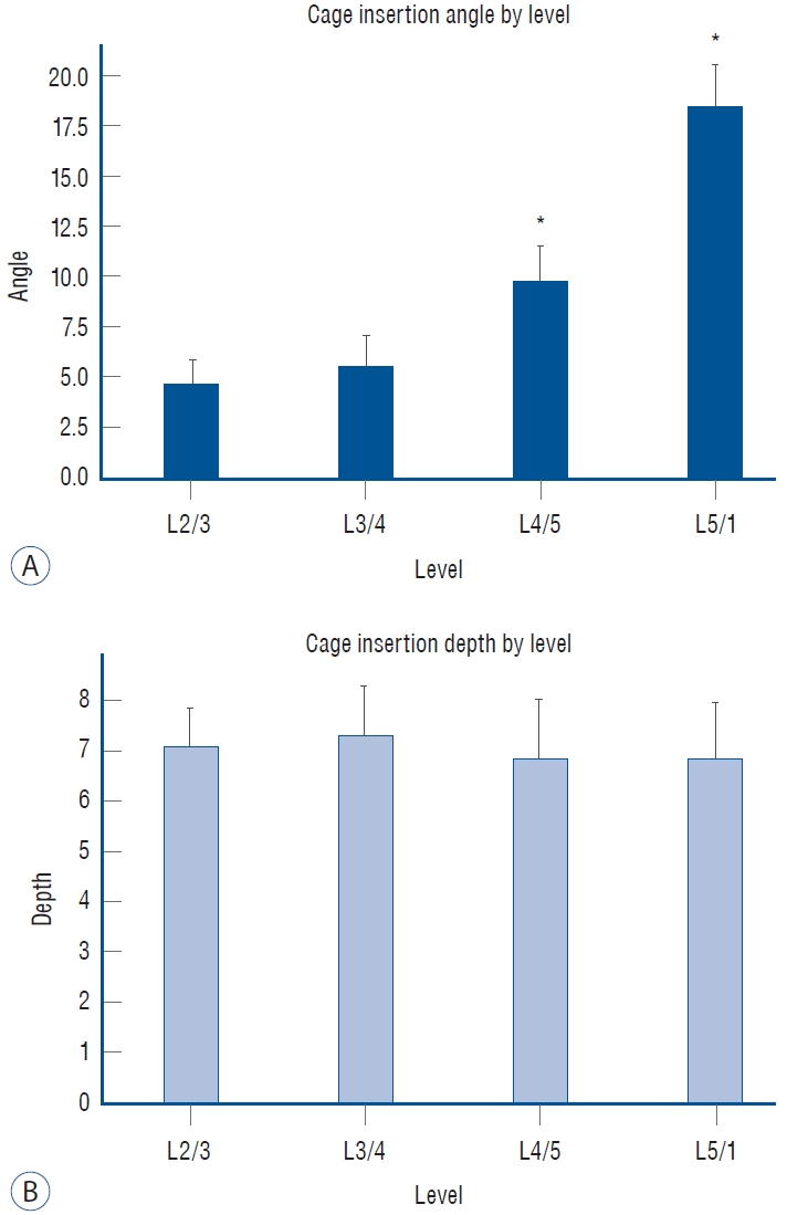
The relationship between cage insertion angle (A) and cage insertion depth (B) according to the level. Values are presented as mean ±standard deviation. *Indicate statistical significance compared to L23.

Cage insertion angle and depth
For preoperative spinal stenosis, 414 (63.7%) of the 650 levels analyzed were confirmed to have spinal stenosis of grade A or B. Of the remaining 236 levels (36.3%), 227 exhibited grade C (96.2%), and nine with grade D (3.8%). Notably, all instances of grade D were at the L4/5 level. Among the nine patients with preoperative grade D, seven (77.8%) improved to postoperative grade A or B, while two (22.2%) transitioned to postoperative grade C, indicating insufficient decompression. Of the 227 patients with preoperative grade C, 218 (96.04%) improved to grades A or B postoperatively, but nine (3.96%) remained at grade C (Table 4). None of the 11 patients with postoperative grade C required additional surgery. When evaluated by level, sufficient indirect compression was achieved at both L2/3 and L5/S1. However, insufficient decompression was found in four cases (1.84%) at L3/4 and in six cases (1.72%) at L4/5.

Pre-operative and post-operative spinal stenosis grade
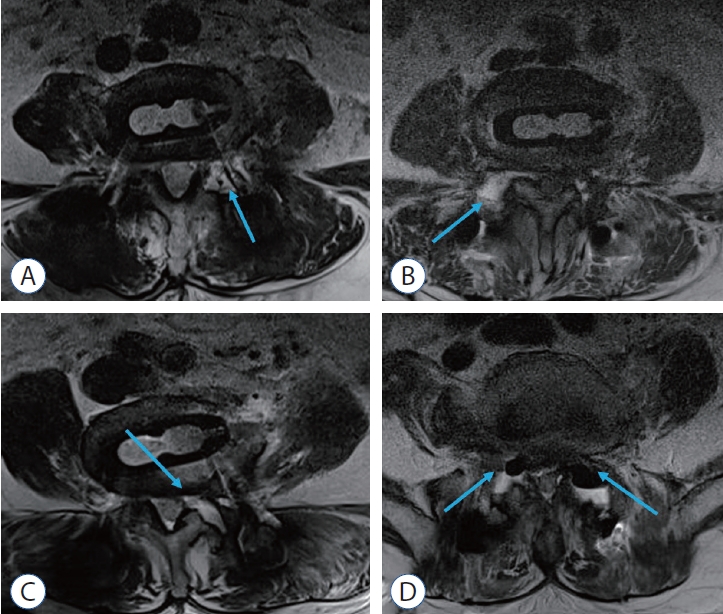
Remnant disc rupture occurred only at the L4/5 and L5/S1 levels. There was one case of central-type disc rupture and three cases of disc rupture towards the contralateral neural foramen. The patient with the central type of disc rupture had previously undergone discectomy, and a new disc rupture occurred at the previous annulotomy site (Fig. 5A). Among the patients with contralateral disc rupture, one patient experienced severe right leg pain postoperatively, and the postoperative MR scan confirmed a disc rupture towards the contralateral neural foramen. This patient had deep cage insertion (distance, 4.5 mm). Even after undergoing revision right facetectomy and neural decompression, the patient consistently reported severe right leg pain (Fig. 5B). The other two patients exhibited symptoms of right foot drop despite not having deep cage insertion. The postoperative MR scans for these patients showed disc rupture towards the right neural foramen. Both patients underwent electromyography/nerve conduction studies, which confirmed radiculopathy, and peroneal palsy was ruled out in the diagnosis. One of them underwent a revision operation, but the recovery was limited to partial muscle strength improvement (MRC grade “zero” to “poor”) (Fig. 5C). The other did not undergo any additional surgery, and there was no recovery in muscle strength up to 1 year postoperatively (MRC grade “zero” to “trace”) (Fig. 5D).

Remnant disc rupture cases. Central disc rupture (A), disc rupture on the contralateral neural foremen, with deep cage insertion (B), disc rupture on the contralateral neural foremen, without deep cage insertion but accompanying right foot drop (C and D). Blue arrows : remnant disc rupture, red arrow : root.
The rupture of a synovial cyst was an unexpected complication. It occurred in four cases and only at the L4/5 level. Notably, facet subluxation was observed preoperatively in all patients. In three cases, manageable lower leg pain was reported (Fig. 6A-C). However, in one case, severe bilateral foot drop occurred postoperatively. Despite performing an emergency subtotal laminectomy, the recovery of muscle strength was limited (MRC grade, right “poor” to “good” left “trace” to “poor”) (Fig. 6D).
Synovial cyst rupture. A-C : Synovium rupture after surgery, but with mild symptoms. D : Synovium rupture and bilateral foot drop after surgery. Blue arrows : synovium rupture.
DISCUSSION
Previous studies on OLIF have focused on symptomatic complications. Among these, the most common complications were psoas paresis, which was reported to be transient [7]. Major complications, such as injuries to the major vessels, ureter, endplate, and sympathetic chain, are also very significant and can be identified during surgery. Postoperative symptoms, such as left thigh weakness due to psoas paresis, decreased temperature sensation in one leg due to sympathetic chain injury, sensory loss and abdominal wall weakening leading to incisional hernia due to genitofemoral nerve damage, can occur due to intraoperative damage [26]. Residual postoperative symptoms can arise from insufficient decompression [24]. The emergence of new symptoms in the opposite leg is difficult to predict, but some case reports have been published [8,16,20]. However, these complication reports are based on intraoperative damage or postoperative symptoms, limiting the identification of potential findings. Especially since postoperative MR scans are usually performed only when symptoms are present, determining the prevalence of such findings is challenging. We conducted MR scans on all patients at 1 week postoperatively starting in December 2019 to identify potential OLIF complications, allowing us to gather ample data on potential complications.
The most common MR finding was approach site hematoma. Despite thorough irrigation and bleeding control after bone cage insertion, we observed hematomas in a surprisingly high proportion of patients (65.3%). However, there were no accompanying symptoms, and it could be considered a subclinical finding due to dead space fluid collection. Nonetheless, caution is needed because the occurrence of hematomas increased when more levels underwent fusion. Although it was excluded from this study, there was a case where a large hematoma after multi-segment surgery transformed into an abscess pocket. Hence, caution is advised for large hematoma occurrences (Supplementary Fig. 1). Another report also showed that a large amount of hematoma led to approach-side symptoms and abscess formation [5,19]. In this context, considering drain insertion at the abdominal approach site when performing fusion on 3 or more levels might be necessary.
Additionally, contralateral psoas hematoma was found in 19% of patients. This is associated with the contralateral annulotomy process, which is essential for inserting a taller and/or wider cage. It mainly occurs when the Cobb elevator is deeply inserted. Fortunately, in our patient group, scanty hematomas were observed, and no patients reported any related symptoms. However, another report showed that when the segmental vessel around the contralateral annulus was damaged, a large hematoma occurred, leading to symptoms in the opposite leg [2].
At the caudal level, we observed that the cage insertion angle tended to be higher and deep cage insertion occurred more frequently, leading to occurrences of cage malposition. This requires careful consideration. Of course, during surgery, we took great care to prevent deep insertion and aimed to maintain the highest degree of orthogonality. However, individual patients have varying pelvic heights, and those with a higher pelvis naturally possess a larger insertion angle [13]. Moreover, for the L5/S1 ante-psoas approach, the cage is inserted at a considerably steep angle. In our study, we identified one patient with mild right leg pain due to deep insertion. Additionally, despite being excluded from the present study (due to the absence of a postoperative MR scan in the initial cases), we reported a revision caused by cage malposition at the L5/S1 level [18]. In this case, the cage malposition resulted from both deep insertion and high-angle insertion. Another risk factor for cage malposition is an incorrect evaluation of the true AP image during surgery, leading to mis-orientation and subsequent deep insertion. It is crucial to recognize that the intraoperative image and the actual cage position can differ significantly, especially with increased obliquity [4]. For safe cage insertion, it is imperative to be cautious when selecting a long and wide cage [9]. It might be preferable to use a smaller cage when a high-angle insertion is expected. In addition, further research is needed on the cage position in intraoperative images compared to its actual position.
Many studies have been conducted regarding indirect decompression. Factors related to insufficient decompression include severe stenosis with high preoperative disc height, impaired preoperative segmental motion, the use of short cages, and severe postoperative subsidence [29]. However, there are still no precise criteria for insufficient indirect decompression. In radiological terms, a study by Shimizu et al. [24] defined cases with grade C and above as insufficient decompression. However, these results are purely radiological and not based on patient symptoms. In our study, we also defined patients with postoperative grade C and above as having insufficient indirect decompression, which was identified in only 4.66% of cases. Fortunately, a chart review showed that most patients with neurogenic intermittent claudication symptoms improved. In this regard, the radiological appearance of grade C stenosis alone is of little significance. Furthermore, in light of reports that the ligamentum flavum can thin over time [15], a decision on additional surgery cannot be made based solely on the finding of insufficient indirect decompression immediately after surgery. In addition, caution is necessary in interpreting these results. This study only involved patients undergoing indirect decompression. We performed direct decompression in cases accompanied by resting pain with severe stenosis, disc rupture, synovial cysts, or lateral bony recess [11]. These indications are similar to those for indirect decompression reported in recent studies [29]. Since these are still low-evidence indications at the level of expert opinion, further research is needed to achieve a consensus.
Remnant disc rupture occurred in four cases. These patients were diagnosed with newly developed disc ruptures based on comparisons with their preoperative MR scans. All cases of disc rupture were accompanied by severe symptoms, such as leg pain and foot drop. Central disc rupture occurred at the site of a previous annulotomy (Fig. 5A). Considering that a significant number of our patients had previously undergone laminectomy, this is considered a rare occurrence. Potential risk factors include a short interval between the previous surgery and the OLIF procedure (within 1 year), and the use of a trial of cage when discectomy was not sufficiently performed during surgery. One case of contralateral neural foramen disc rupture was associated with deep cage insertion (Fig. 5B). In this case, the distance from the cage to the neural foramen was relatively close (4.5 mm). Hence, we set the threshold for deep insertion at 5 mm. While cage malposition or deep cage insertion is something we can anticipate and adjust for intraoperatively, the other cases (Fig. 5C and D) were unpredictable, as remnant disc rupture occurred even though the cages were not deeply inserted and the cage insertion angle was not large. Although the incidence rate of this complication is low, making it challenging to determine the exact cause, we believe the most likely reason is that sufficient discectomy was not performed before using the trial, and the remnant disc material was pushed by the trial, causing the disc to rupture at the weak contralateral annular corner site. Fully understanding the underlying factors is a complicated task, necessitating more in-depth research in the future. At present, the best practices to prevent remnant disc rupture include avoiding deep insertion when there is insufficient orthogonality and emphasizing the importance of adequate discectomy prior to trial use.
The rupture of a synovial cyst was an entirely unexpected complication. Typically, facet joint widening is also used as evidence of good indirect decompression [27]. However, rare symptoms due to the rupture of the synovium were identified in four cases. Among them, three cases had mild symptoms that could be treated with medication (Fig. 6A-C), but one patient had bilateral foot drop, requiring us to be especially cautious (Fig. 6D). All four patients had preoperative facet subluxation. However, despite a substantial number of enrolled patients in our study presenting with spondylolisthesis or facet subluxation, synovium rupture occurred in only a very small subset of these patients. In this regard, synovium rupture does not necessarily occur just because there is subluxation, but in some patients with subluxation, synovium rupture can occur after OLIF. It is expected that additional risk factor analysis will be possible as cases accumulate in the future. In a literature review, the study by Parikh and Jhala [21] described a remnant disc, but their finding is thought to have resulted from actual synovium rupture. This patient also had facet subluxation. As presented in Fig. 6C, the natural absorption of the synovium rupture was confirmed on the MR scan after 3 months, but the mild symptoms persisted up to 1 year after surgery. We have identified facet synovium rupture as a novel complication that can cause new symptoms or the persistence of existing symptoms after OLIF surgery. Additionally, we recommend that future MR analyses should include an evaluation of this possible complication.
Limitations
This study excluded patients who underwent laminectomy, which was additionally performed in cases of severe stenosis, lateral recess, disc rupture, and symptomatic synovial cyst. Laminectomy was often performed to decompress a specific level in patients with multi-level disease. In this regard, there is an inherent selection bias since the findings of this study do not reflect all OLIF patients. However, since remnant disc rupture, synovium rupture, and other complications cannot be confirmed when laminectomy is performed, we believe that our research provides novel information about OLIF, the benefits of which mainly derive from indirect decompression. The presence of numerous subtle symptoms among the clinical outcomes could also cast doubt on the objectivity of clinical assessments in our study. However, since we have been meticulously collecting complications since the start of OLIF at our institution [14,18,27], we believe that we can resolve the occurrence of residual or new symptoms, as we have accumulated extensive data prospectively. Lastly, there were few cases of clinically important symptomatic complications such as remnant disc rupture or synovial cyst rupture. Therefore, it was difficult to analyze the risk factors for these conditions.
CONCLUSION
Our study on OLIF complications reveals that approach site and contralateral psoas hematomas are common but typically not clinically significant. We observed that cage malposition, associated with decreased orthogonality and deeper insertion at lower levels, requires attention during surgery. Notably, only a small percentage of patients experienced incomplete indirect decompression, which did not necessitate additional surgery. While remnant disc rupture and synovium rupture were identified as serious complications, they occurred infrequently. These findings underscore the importance of careful intraoperative technique and highlight the utility of postoperative MR scans in detecting potential complications associated with OLIF.
Notes
Conflicts of interest
Dong-Wuk Son has been editorial board of JKNS since November 2017. He was not involved in the review process of this original article. No potential conflict of interest relevant to this article was reported.
Informed consent
This type of study does not require informed consent.
Author contributions
Conceptualization : KHL, SHL, DWS; Data curation : KHL, SHL, JSL, DWS; Formal analysis : KHL, SHL, YHK; Funding acquisition : SHL; Methodology : KHL, SHL, JSL, DWS; Project administration : KHL, SHL; Visualization : KHL, SHL; Writing - original draft : KHL, SHL; Writing - review & editing : KHL, SHL, JSL, YHK, SKS, DWS, SWL, GSS
Data sharing
None
Preprint
None
Acknowledgements
This study was supported by Research institute for Convergence of biomedical science and technology (30-2023-006), Pusan National University Yangsan Hospital.
Supplementary materials
The online-only data supplement is available with this article at https://doi.org/10.3340/jkns.2023.0238.
Transformation of approach site hematoma into abscess. A 78-year-old woman underwent 4-level OLIF (L2/3, 3/4, 4/5, 5/1) and decompressive laminectomy at L2 and L5. One week postoperatively, an magnetic resonance (MR) scan revealed a significant amount of hematoma (A and B). At 4 weeks postoperatively, due to elevated inflammatory markers, abdominal computed tomography (CT) was performed, identifying an abscess approximately 8 cm in size (C and D). Percutaneous drainage was conducted, followed by 4 weeks of intravenous antibiotic therapy and 2 weeks of oral antibiotics. A and B : Postoperative MR scans showing a large amount of hematoma (blue arrows). C and D : CT of the abdomen with contrast enhancement, showing the formation of an abscess (red arrow) at the approach site.