| Home | E-Submission | Sitemap | Editorial Office |  
top_img
Journal of Korean Neurosurgical Society > Volume 66(2); 2023 > Article
Maoneo, Beltchika, Kabulo, and Ntsambi: Letter to the Editor : Extramedullary Intradural Calcified Meningiomas Surgery in Limited Resources Context
To the Editor,
Spinal meningiomas are benign, slow-growing tumours, developed from arachnoidal cap cells [4]. Calcified forms are about 1-5% of all meningiomas [3]. Complete surgical removal of calcified spinal meningioma remains a true challenge in a context of limited resources without a neurosurgical microscope because of possible dural sac and nerve roots adhesions [3,4]. We write this letter to the editor with the aim to explain our critical circumstances of the work according to three patients.
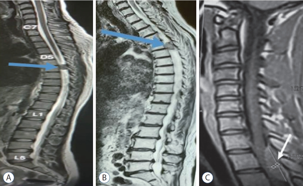
We experienced three cases of calcified spinal meningiomas, the clinical features are summarized in Table 1. Fig. 1 shows the magnetic images taken before the operation.
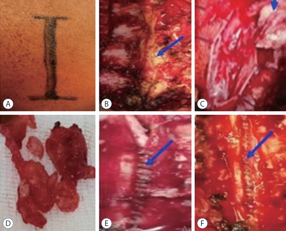
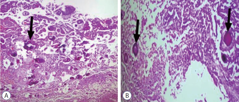
In all three cases, our surgical procedures consisted of (Fig. 2) : 1) identification of the level using the MRI or CT-scan images. We performed laminectomy of two or three levels depending on the extent of the tumor on the imaging. 2) Complete release of the dural sac posteriorly and postero-laterally. 3) Instrumental palpation of the tumor mass to assess the consistency. 4) Then longitudinal durotomy of about 3-4 cm of the external dural layer using micro-instruments, going beyond the tumour region on either side. 5) Gradual dissection of the tumor by removing the adherent internal dural layer while meticulously separating the rootlets attached to the mass according to Saito et al. [6] techniques using micro-instruments. 6) After complete resection of the tumor, control of hemostasis using surgicel followed by watertight closure of the external dural layer using 3/0 vicryl with no use of biological glue. And 8) porto vac drain was left in situ then suture by layer. No dural defects were observed apart from the surgical opening. The specimen taken and sent for histology came back as psammomatous (calcified) meningioma (Fig. 3).
Calcified spinal meningiomas are frequently observed in women over 50 years old, located in the thoracic spine. However, they are also found in people under 50 years of age usually associated with a poor prognosis [2]. The signs can be confused with other diseases in elderly people and delay the diagnosis [1]. The delay in diagnosis is also due to the unavailability and accessibility of CT scan and MRI in countries with limited resources such as ours. Complete removal of the tumour, according to Simpson’s grades I, II, and III, is the best treatment for spinal meningioma. But the risk of cerebrospinal fluid leakage and recurrence is high [5]. The novel technique, similar to Simpson II, developed by Saito and colleagues [6] that consists of removing the meningioma with the inner layer of the dura and keeping the superficial layer for closure has got excellent results [7].
Many neurosurgeons use a surgical microscope during the operation. However, in a context of limited resources, this valuable tool is not always available. In the cases reported, we performed a macroscopically complete removal of the mass with the medial layer of the dura according to the procedure of Saito et al. [6] without neurosurgical microscope.
These cases show the possibility of a complete recovery after a total macroscopic resection, even in a less elderly patient by using Saito technique without a neurosurgical microscope in limited resource context and the risk of the comorbidity in an elderly person.

Notes

Conflicts of interest

No potential conflict of interest relevant to this article was reported.

Informed consent

This type of study does not require informed consent.

Author contributions

Conceptualization : IM; Data curation : IM; Formal analysis : IM, KDMK, GN; Methodology : GN; Project administration : AB, GN; Visualization : KDMK; Writing - original draft : IM; Writing - review & editing: IM, KDMK, GN

Data sharing

None

Preprint

None

Fig. 1.
Preoperative sagittal magnetic resonance images of case 1 (A), 2 (B) and 3 (C). T2 weighted image shows an intradural and extramedullary meningioma (blue arrow in A, B; white arrow in C).
jkns-2022-0125f1.jpg
Fig. 2.
Intraoperative images showing. A : The posterior approach. B : The spinous processes resection and laminectomy (blue arrow). C : The dural opening through a longitudinal incision and the detachment and removal of the meningioma with the internal layer according to Saito et al.6). D : The meningioma mass resected. E and F : A watertight suture of the dural superficial sheet (blue arrow).
jkns-2022-0125f2.jpg
Fig. 3.
A and B: Histopathology picture shows numerous calcification foci (psammomas) (black arrow) (A : Hematoxylin and Eosin [H&E] staining, ×10; B : H&E staining, ×40).
jkns-2022-0125f3.jpg
Table 1.
Clinical Summary of three patients
Case No. Age (years)/sex Location of tumor Neurological findings Treatment outcome
1 29/F T7 Paraparesis, sensory deficit, hyperactive DTR Total removal, good recovery
2 69/F T5, 6 Paraparesis Total removal, death due to medical problem
3 20/F T2, 3 Paraparesis, sphincter change Total removal, good recovery

F : female, DTR : deep tendon reflex

References

1. Apostolov G, Kehayov I, Kitov B : Clinical aspects of spinal meningiomas: a review. Folia Med (Plovdiv) 63 : 24-29, 2021
pmid
2. Cohen-Gadol AA, Zikel OM, Koch CA, Scheithauer BW, Krauss WE : Spinal meningiomas in patients younger than 50 years of age: a 21-year experience. J Neurosurg 98(3 Suppl):258-263, 2003
crossref pmid
3. Kim JY, Min WK, Kim JE, Park KH, Mun JU : Two case reports of calcified spinal meningioma and a literature review. J Korean Soc Spine Surg 23 : 227-233, 2016
crossref pdf
4. Kobayashi K, Ando K, Matsumoto T, Sato K, Kato F, Kanemura T, et al : Clinical features and prognostic factors in spinal meningioma surgery from a multicenter study. Sci Rep 11 : 11630, 2021
crossref pmid pmc pdf
5. Miyakoshi N, Hongo M, Kasukawa Y, Shimada Y : En-bloc resection of thoracic calcified meningioma with inner dural layer in recapping T-saw laminoplasty: a case report. BMC Surg 15 : 82, 2015
crossref pmid pmc pdf
6. Saito T, Arizono T, Maeda T, Terada K, Iwamoto Y : A novel technique for surgical resection of spinal meningioma. Spine (Phila Pa 1976) 26 : 1805-1808, 2001
crossref pmid
7. Tominaga H, Kawamura I, Ijiri K, Yone K, Taniguchi N : Surgical results of the resection of spinal meningioma with the inner layer of dura more than 10 years after surgery. Sci Rep 11 : 4050, 2021
crossref pmid pmc pdf
TOOLS
PDF Links  PDF Links
PubReader  PubReader
ePub Link  ePub Link
XML Download  XML Download
Full text via DOI  Full text via DOI
Download Citation  Download Citation
  Print
Share:      
METRICS
0
Crossref
0
Scopus
1,695
View
56
Download
Related article
Editorial Office
1F, 18, Heolleung-ro 569-gil, Gangnam-gu, Seoul, Republic of Korea
TEL: +82-2-525-7552   FAX: +82-2-525-7554   E-mail: office@jkns.or.kr
About |  Browse Articles |  Current Issue |  For Authors and Reviewers
Copyright © Korean Neurosurgical Society.                 Developed in M2PI
Close layer